


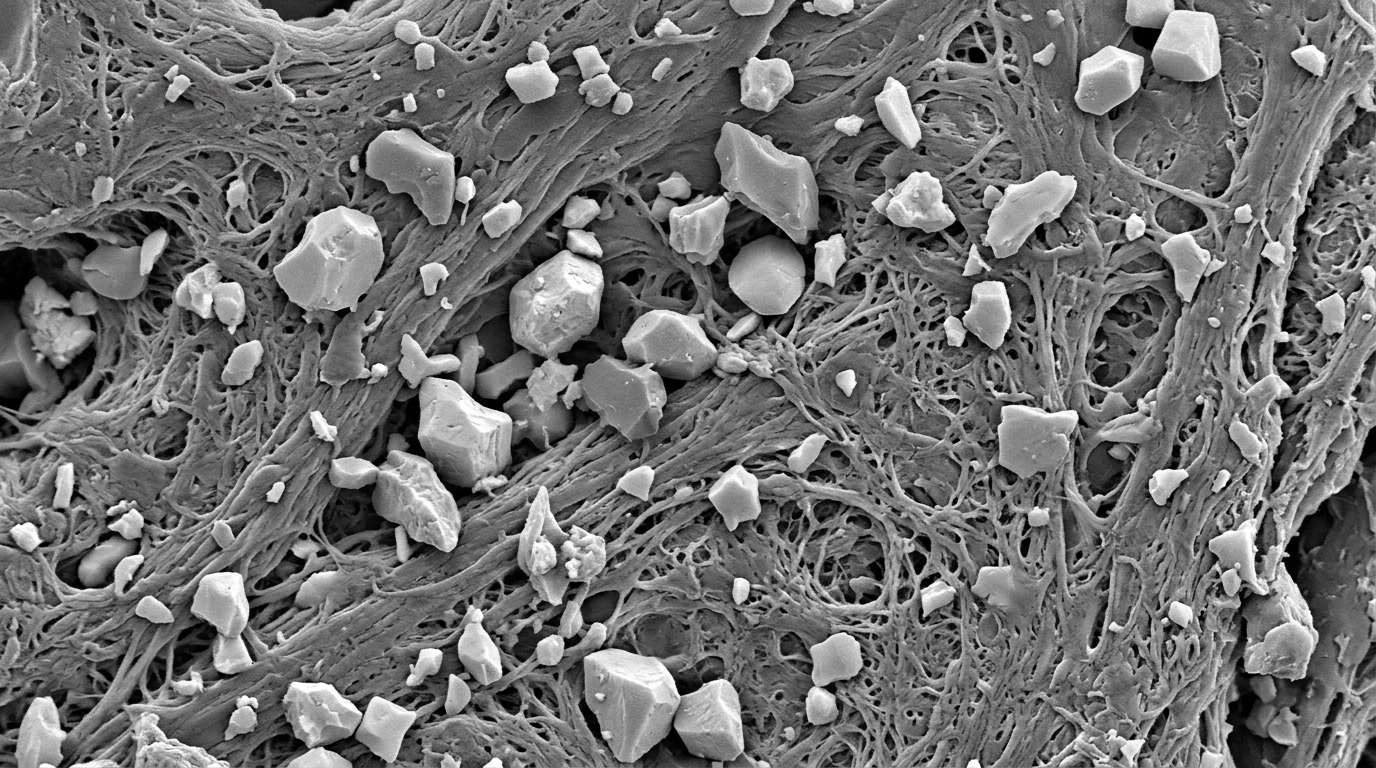
Published in Applied Microscopy, the study presents a general protocol designed to characterize micro- and nano-structural features of debris while it remains partially embedded in surrounding biological tissue. This approach addresses a significant gap in current laboratory practices, where debris is typically fully isolated from tissue before analysis, a process that can alter particle morphology and obscure agglomeration phenomena within the body.
The primary objective of the research is to provide a reliable modus operandi for both research and hospital laboratories. By following a proven methodology, these institutions can reduce analysis time and obtain results associated with a common protocol, leading to more comparable data across different studies. The methodology was developed and validated using a specific clinical case: a hip prosthetic revision where a chromium-cobalt (Cr-Co) femoral head misalignment caused the wearing out of the polyethylene acetabular insert and partial wear of the titanium-6-aluminum-4-vanadium (Ti-6Al-4 V) acetabular cup.
The study focuses on the release of prosthesis debris at the tissue-implant interface, a major cause of aseptic loosening. Aseptic loosening is a phenomenon that often requires the premature replacement of the prosthesis. The authors note that biophysical phenomena at the interface between prosthetic components and surrounding tissues strongly determine the success or failure of arthroplasty surgery. Causes for the generation of micro-nanometric fragments include partial osseointegration, tribo-mechanical factors such as micro-displacement, and the aging process of prosthetic surfaces associated with corrosion and wear.
Previous studies have largely focused on isolating particulate matter completely from tissue to allow for in-depth characterization and better 2D and 3D imaging. However, this complete isolation can be detrimental to the size, shape, and number of particles, potentially altering their morphology or causing them to agglomerate. The proposed protocol in this study allows for the study of wear fragments still partially embedded in the tissue. While this results in a less complete characterization of individual fragments compared to fully isolated samples, it affords the possibility of evaluating how biological tissues respond to the presence of debris and assessing agglomeration effects occurring within the tissue.
The experimental design involved biopsy samples taken during hip, shoulder, and knee prosthetic revision surgeries in three Italian hospitals. The specific specimen used for validation was from a clinical case involving an uncemented left hip prosthesis that failed after 17 years due to the decentralization of the Cr-Co femoral head relative to the Ti-6Al-4 V cup. This decentering was diagnosed via pre-explant radiography and was responsible for the wear of prosthesis components, leading to osteolysis and aseptic loosening. The samples consisted mainly of foreign body granulomas extracted from the joint pseudo-capsule, which contained a large amount of metal and polyethylene debris.
For sample preparation, biopsy samples embedded in kerosene were microtome sectioned into slices approximately 2 micrometers thick. The researchers utilized a partial digestion technique using potassium hydroxide (KOH) at concentrations ranging from 0.2 M to 2 M for one hour. This digestion process was chosen because it did not allow for the complete isolation of particulate matter, thereby preserving the opportunity to observe particulate agglomeration phenomena. The authors noted that higher concentrations or longer digestion times, as suggested by some previous literature, could change the size of metal particles and jeopardize morphological and quantitative analysis. The partially digested tissues were then gold-coated to prevent electrical charging during SEM analysis.
The methodology follows a hierarchical, stepwise approach. The first step involves low-magnification SEM observation using both secondary electron (SE) and backscattered electron (BSE) images to identify areas of interest. BSE images are particularly useful for detecting metal particles, which appear as bright spots on a dark gray background due to their higher atomic number compared to the organic tissue matrix. Once a suitable area is identified, a full-field EDX spectral analysis is conducted to identify the elements present, primarily titanium, aluminum, vanadium, iron, cobalt, and chromium.
Following the spectral analysis, full-field EDX elemental mapping is performed to locate the elements of interest within the sample. The researchers set a threshold for normalized elemental concentrations higher than 1 wt% to distinguish relevant debris from surface contamination. Subsequent high-magnification SEM observations allow for the detailed investigation of particle morphology, surface topography, and size. In this specific case, the EDX analysis confirmed that the debris originated from the prosthetic components, showing titanium, aluminum, and vanadium from the acetabular cup, and trace amounts of cobalt and chromium from the femoral head.
The high-magnification analysis revealed that what appeared as single particles at lower magnifications were actually clusters of agglomerated particles within the periprosthetic tissue. The researchers measured the aspect ratio of 45 titanium alloy particles, defined as the ratio of the major axis to the minor axis. The calculated aspect ratio was 2.5 ± 1.2, with a range of 1.2 to 7.3. This value indicates that the particles were rather elongated, though not to the point of being fibrillar. The morphology was described as irregular, granular, and in some cases plate or shard-like, consistent with ASTM F1877 standards for wear debris.
The findings regarding particle size and morphology are compatible with previous studies, such as those by Topolovec et al. and Stratton Powell et al., which also observed titanium alloy particles with similar aspect ratios in periprosthetic tissues. However, this study differs in its in situ analysis approach, which allowed for the observation of clustering effects that were not as prominent in studies involving fully digested samples. The authors hypothesize that the tribological origin of the debris is due to two main mechanisms: an adhesive mechanism between the softer titanium alloy and the harder chromium-cobalt alloy, and a fretting wear process associated with delamination of the titanium alloy.
The study highlights the importance of understanding the specific wear processes involved in prosthetic failure, such as sliding wear and alternating sliding wear. These processes are caused by the geometry of the wear paths and the size of the contact area relative to the stroke of the wear path. Adhesion, abrasion, surface fatigue, and tribochemical reactions are often present simultaneously. While titanium alloys are known for their corrosion resistance due to the formation of a stable TiO2 passive layer, they are not immune to wear, particularly over long periods. The safety of aluminum and vanadium in these alloys is also a concern, as they can be released during the wear process.
The proposed protocol is intended to be a guide for laboratories needing a rigorous and tested modus operandi. It is designed to be easily adoptable to similar samples, optimizing the analysis procedure and reducing test time, which is a prerequisite for managing the massive volume of samples often encountered in hospital contexts. The critical discussion of the results clarifies the applicability fields of the methodology, noting that while it was developed for metallic debris, it can be adapted for other materials involved in arthroplasty, such as polymeric and ceramic components.
This research contributes to the broader goal of developing more biocompatible implants with greater longevity. As the number of joint replacements in younger patients increases and the life expectancy of the elderly extends, understanding the causes of prosthetic failure becomes increasingly critical. By providing a standardized method for analyzing intra-tissue wear debris, the study aims to facilitate future research into tribological phenomena and the biological response to prosthetic implants, ultimately helping to improve the outcomes of arthroplasty surgeries.
Subject of Research: From a clinical case to a general methodology to analyze prosthetic joint failure, by micro- and nano-characterization with SEM of intra-tissue wear debris
Article Title: From a clinical case to a general methodology to analyze prosthetic joint failure, by micro- and nano-characterization with SEM of intra-tissue wear debris
Article References: Tarter, S., Maines, L., Nollo, G., Molinari, M., & Gialanella, S. (2025). From a clinical case to a general methodology to analyze prosthetic joint failure, by micro- and nano-characterization with SEM of intra-tissue wear debris. Applied Microscopy, 55(1), Article 14. https://doi.org/10.1186/s42649-025-00118-2
Image Credits: AI Generated
DOI: 10.1186/s42649-025-00118-2
Keywords: clinical, case, general, methodology, analyze, prosthetic, joint, failure, micro-, nano-characterization, intra-tissue, wear
Cite Scienmag News
Denise Maddox. (November 8, 2026). New SEM Protocol Maps Wear Debris Inside Joint Tissue. Scienmag. https://scienmag.com/new-sem-protocol-maps-wear-debris-inside-joint-tissue/
Denise Maddox. "New SEM Protocol Maps Wear Debris Inside Joint Tissue." Scienmag, 8 November 2026, https://scienmag.com/new-sem-protocol-maps-wear-debris-inside-joint-tissue/. Accessed 10 November 2026.
Denise Maddox. "New SEM Protocol Maps Wear Debris Inside Joint Tissue." Scienmag. November 8, 2026. https://scienmag.com/new-sem-protocol-maps-wear-debris-inside-joint-tissue/




