


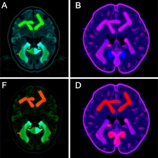
A new neonatal model of hypoxic-ischemic injury is challenging a central assumption in the study of newborn brain damage: that lasting cognitive problems must be accompanied by obvious neuronal loss. In a study published in Pediatric Research, Langer, Tiemeier, Harmon and colleagues report that their model produced persistent glial activation and memory deficits even though they did not detect widespread loss of neurons. The findings suggest that early-life oxygen deprivation and reduced blood flow may disrupt brain function through cellular and inflammatory changes that remain hidden when researchers focus primarily on neuronal death.
Hypoxic-ischemic injury occurs when the brain receives too little oxygen and insufficient blood supply. In newborns, it can follow complications such as birth asphyxia, placental or umbilical cord problems, respiratory failure, or severe cardiovascular instability. The condition can trigger a rapidly evolving cascade: energy production collapses, ion gradients fail, excitatory neurotransmitters accumulate, calcium floods into cells, and oxidative stress damages membranes and organelles. In severe cases, neurons die within hours or days. Yet the new work indicates that injury can also leave behind a more subtle biological signature, in which neurons survive but the networks supporting learning and memory do not function normally.
The researchers developed a neonatal hypoxic-ischemic paradigm intended to capture aspects of injury that may be missed by conventional models. Rather than treating neuronal survival as the definitive measure of recovery, the study combined behavioral testing with cellular and tissue-level analyses. The model allowed the team to examine how early oxygen deprivation affects later cognition, while also determining whether the brain showed signs of inflammation or structural neuronal loss. This combined approach is important because a brain can retain its neurons and still suffer from impaired synaptic communication, altered circuit development, or persistent immune activity.
One of the study’s most notable observations involved glial cells, the non-neuronal cells that support, protect, and regulate the nervous system. Microglia act as the brain’s resident immune cells, surveying tissue and responding to danger signals. Astrocytes maintain the chemical environment around neurons, help regulate neurotransmitters and ions, and contribute to the blood-brain barrier. After injury, both cell types can change shape, gene expression, and behavior. These responses can be beneficial at first, helping to clear damaged material and stabilize the tissue, but prolonged activation may interfere with synapse formation, alter neuronal signaling, and sustain a damaging inflammatory environment.
In the neonatal model, markers of glial activation were present even without detectable neuronal loss in the examined regions. That result points to a form of brain dysfunction driven less by the disappearance of neurons than by changes in the conditions in which neurons operate. Activated microglia can release cytokines and other signaling molecules that influence synaptic plasticity, while reactive astrocytes may alter glutamate uptake, energy support, and extracellular ion balance. During early development, when neural circuits are being assembled and refined, these disturbances could have consequences that emerge only later, when the animal is required to learn, remember, and adapt.
The behavioral findings provided that delayed signal. Animals exposed to the neonatal insult later showed deficits on memory-related tasks, according to the study, despite the absence of measurable neuronal loss. Such tests are commonly used to assess the ability to learn associations, remember locations or contexts, and distinguish familiar from novel information. Performance depends on coordinated activity across networks that include the hippocampus and connected cortical regions. A memory deficit therefore does not necessarily indicate that one isolated structure has been destroyed; it can reflect altered synaptic strength, impaired circuit connectivity, abnormal neuroinflammatory signaling, or disrupted maturation of the brain’s communication systems.
This distinction could help explain why some children affected by neonatal hypoxic-ischemic events experience cognitive, attention, or learning difficulties even when standard imaging does not show extensive tissue destruction. Clinical scans are valuable for identifying major injury, but subtle cellular dysfunction may fall below their resolution. The study’s findings raise the possibility that glial activity and related molecular changes could serve as earlier or more sensitive indicators of risk. If validated in additional models and eventually in patients, such biomarkers might help clinicians identify infants who need long-term developmental monitoring even when overt neuronal damage appears limited.
The work also has implications for treatment. Current management of moderate to severe neonatal hypoxic-ischemic encephalopathy can include therapeutic hypothermia, which lowers metabolic demand and can reduce the extent of injury when delivered within a defined clinical window. However, cooling is not universally effective, and it does not eliminate the possibility of later cognitive or behavioral problems. A model in which glial activation persists without neuronal death could be useful for testing therapies aimed at neuroinflammation, microglial state, astrocyte function, synaptic plasticity, or metabolic recovery. The goal would not simply be to prevent neurons from dying, but to preserve the quality of the circuits those neurons form.
The study does not mean that neuronal loss is unimportant or that all neonatal hypoxic-ischemic injuries follow the same pattern. The severity, timing, duration, and developmental stage of an insult can produce very different outcomes. In addition, behavioral effects in an experimental model cannot be translated directly into predictions for individual infants. The reported absence of neuronal loss also depends on the brain regions examined, the markers used, and the time points selected for analysis. Further research will be needed to determine how long glial activation persists, which molecular pathways drive the memory deficits, and whether reversing those changes can restore normal cognitive performance.
By separating neuronal survival from functional recovery, Langer and colleagues offer a more nuanced picture of neonatal brain injury. The results suggest that the developing brain may retain its basic cellular architecture while undergoing changes that weaken communication between cells and impair the formation of durable memories. That possibility could shift the field toward a broader definition of brain protection—one that measures not only how many neurons remain, but also how effectively neurons, glia, and neural circuits work together after an early-life insult.
Cite Scienmag News
Harold Sullivan. (August 3, 2026). New neonatal hypoxic-ischemic model shows glial activation, memory deficits without neuronal loss. Scienmag. https://scienmag.com/new-neonatal-hypoxic-ischemic-model-shows-glial-activation-memory-deficits-without-neuronal-loss/
Harold Sullivan. "New neonatal hypoxic-ischemic model shows glial activation, memory deficits without neuronal loss." Scienmag, 3 August 2026, https://scienmag.com/new-neonatal-hypoxic-ischemic-model-shows-glial-activation-memory-deficits-without-neuronal-loss/. Accessed 3 September 2026.
Harold Sullivan. "New neonatal hypoxic-ischemic model shows glial activation, memory deficits without neuronal loss." Scienmag. August 3, 2026. https://scienmag.com/new-neonatal-hypoxic-ischemic-model-shows-glial-activation-memory-deficits-without-neuronal-loss/


