A groundbreaking advancement in the imaging and surgical treatment of osteosarcoma promises to revolutionize how this aggressive bone cancer is managed, offering hope for improved outcomes through cutting-edge precision technology. At the upcoming Society of Nuclear Medicine and Molecular Imaging (SNMMI) 2026 Annual Meeting, researchers from Peking University Cancer Hospital and Institute in Beijing will unveil an innovative integrated PET imaging platform capable of rapidly and accurately distinguishing malignant tumor tissue from healthy tissue during surgery. This novel system not only enhances real-time decision-making in the operating room but also enables precise assessment of surgical margins, a critical factor in fully eradicating tumors and minimizing recurrence.
Osteosarcoma, the most common primary malignant bone tumor affecting children and adolescents, represents a formidable clinical challenge. The current therapeutic standard combines aggressive chemotherapy with radical surgical excision. A paramount objective for surgeons is to remove the entire tumor with clear margins, as residual tumor cells within resection boundaries markedly increase the risk of local recurrence and negatively impact patient survival. However, delineating tumor margins intraoperatively with confidence remains difficult, frequently requiring surgeons to make empirical decisions based on visual and tactile feedback—methods insufficient for microscopic precision.
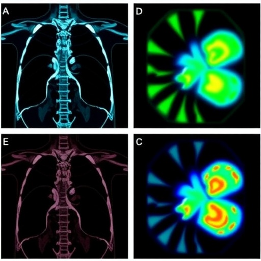
This clinical necessity spurred the development of a sophisticated multi-modal imaging platform engineered to overcome existing limitations. Central to the technology is the targeting of B7-H3, a transmembrane protein highly expressed in over 80% of osteosarcoma tumors. Recognizing this protein’s selective overexpression, researchers successfully synthesized a novel radiotracer, designated ^68Ga-B7H3-BCH, that selectively binds to B7-H3 molecules, enabling highly specific and sensitive detection of osteosarcoma lesions through PET imaging.
Preclinical assessments underscored the superior diagnostic capability of the ^68Ga-B7H3-BCH tracer, demonstrating marked improvements in lesion detection and tumor delineation compared to conventional radiotracers like ^18F-FDG. Encouraged by these findings, the research team architected an integrated imaging pipeline that synergizes ^68Ga-B7H3-BCH PET/CT scanning with a near-infrared (NIR) B7H3 fluorescent probe. This dual-modality approach facilitates comprehensive preoperative tumor staging and equips surgeons with real-time fluorescence visualization during tumor resection procedures.
During the surgical phase, the NIR fluorescent probe illuminates tumor borders with high spatial and temporal resolution, guiding surgeons to excise malignant tissues precisely while preserving as much healthy bone and surrounding structures as possible, which is vital for maintaining limb functionality. Following tumor removal, the platform incorporates a rapid pathological margin verification technique capable of providing conclusive margin status within 30 minutes, dramatically expediting what traditionally is a protracted pathological process and enhancing surgical confidence.
Mouse model studies exhibited robust uptake of the ^68Ga-B7H3-BCH tracer within osteosarcoma lesions and at tumor margins, correlating well with histopathological analysis and validating the tracer’s specificity. The combination of non-invasive, whole-body PET/CT imaging for systemic staging and intraoperative fluorescence for margin delineation embodies a truly personalized, closed-loop diagnostic and therapeutic strategy.
The implications of this integrated platform extend beyond mere imaging enhancements. It introduces a paradigm shift toward precision oncology in osteosarcoma, transitioning from empirical surgery followed by standard systemic chemotherapy to individualized treatment plans shaped by precise molecular and anatomical tumor information. Such tailoring is poised not only to improve local control rates but also to reduce unnecessary removal of healthy tissue, ultimately translating into better functional outcomes and quality of life for patients.
Bo Mei, PhD, the principal investigator spearheading this innovation, emphasized the urgent clinical need: “Orthopedic surgeons need a reliable, rapid method to accurately delineate tumor margins in real-time during osteosarcoma surgeries. Our integrated platform meets this challenge, redefining surgical oncology practices by incorporating molecular targeting and advanced imaging modalities.”
Although still at the investigational stage, early human feasibility studies employing the ^68Ga-B7H3-BCH platform have shown promising results. These pilot data demonstrate the platform’s potential to function effectively in clinical settings, marking a critical step toward regulatory approval and widespread adoption. Future efforts will focus on comprehensive prospective clinical trials to robustly establish safety, efficacy, and workflow integration within orthopedic oncology centers.
The technical innovation rests heavily on multimodal probe development, marrying the quantitative power of PET imaging with the exquisite real-time spatial resolution of fluorescence imaging. This combination overcomes intrinsic limitations of each modality when used in isolation—PET provides metabolic and molecular insights but is limited in spatial resolution and intraoperative applicability, while fluorescence enables visual guidance but lacks systemic diagnostic capability.
The platform’s rapid intraoperative margin assessment, with results available in less than half an hour, is a significant advance that replaces delayed histopathology consultation, allowing surgeons to adjust the extent of resection dynamically and immediately. By integrating molecular targeting, imaging, and pathology, this closed-loop diagnostic and therapeutic construct exemplifies next-generation precision medicine and theranostics.
This innovation also represents a promising template for other solid tumors exhibiting targetable biomarkers, suggesting broad applicability across oncology. The integration of molecularly specific PET tracers with intraoperative fluorescence guidance and rapid pathology verification embodies a comprehensive approach that can be adapted and refined for diverse malignancies beyond osteosarcoma.
As SNMMI 2026 unfolds, this pioneering work will undoubtedly attract attention from the nuclear medicine, surgical oncology, and molecular imaging communities. The researchers’ abstract, detailing the development and validation of the ^68Ga-B7H3-BCH PET/fluorescence multimodal probe and integrated imaging platform, underscores the convergence of technology and translational science, poised to enhance patient care profoundly.
This new frontier in osteosarcoma management showcases how targeted molecular imaging coupled with innovative surgical navigation can dramatically improve diagnostic accuracy, surgical precision, and ultimately patient prognosis. It exemplifies the power of integrating molecular biology, chemistry, imaging technology, and clinical expertise into a cohesive solution designed to address one of the most challenging pediatric cancers.
Subject of Research: Osteosarcoma, molecular imaging, surgical margin assessment, precision oncology
Article Title: An Integrated PET Imaging Platform for Real-Time Surgical Guidance and Accurate Margin Assessment in Osteosarcoma
News Publication Date: 2026 (presented at SNMMI Annual Meeting)
Web References: SNMMI 2026 Annual Meeting Abstract
Image Credits: Courtesy of SNMMI
Keywords: Osteosarcoma, B7-H3, PET Imaging, Molecular Imaging, Near-Infrared Fluorescence, Surgical Navigation, Radiotracer, Precision Medicine, Tumor Margin, Theranostics
