


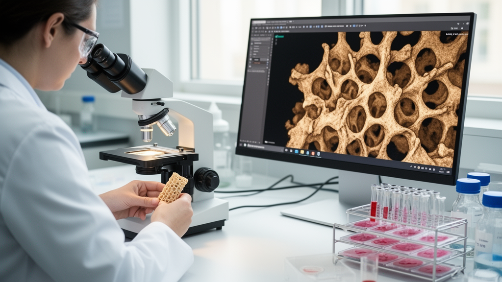
Bone regeneration research has long struggled with a methodological dilemma: the techniques that best reveal the microstructure of healing bone are not the ones that best reveal its biology. A team of Brazilian researchers has now published a comprehensive methodological guideline in Frontiers in Imaging that bridges this gap, pairing X-ray microcomputed tomography (micro-CT) with classical histology in a single standardized workflow for evaluating defect repair in rodent models. By focusing on two anatomically distinct targets—the rat tibia as a long bone and the mouse calvaria as a flat bone—the protocol offers the bone repair community a reproducible framework for assessing both mineralized microarchitecture and the cellular events that drive it.
The clinical stakes are considerable. Bone injuries can severely affect quality of life and productivity while driving up public health spending, and advances in tissue engineering have intensified the search for regenerative strategies and biomaterials. Translational studies typically rely on rodent lesion models, most commonly created with a surgical drill coupled to a micromotor, which produces well-defined circular osteotomies. Compared with fracture models, drilled defects offer clear delimitation and standardized lesion size, yielding statistically representative and reproducible results without requiring animal immobilization—a practical advantage that underpins the entire protocol.
The biology of repair differs fundamentally between the two bone types, which is why the guideline treats them separately. Long bone defects heal through endochondral ossification, with cartilage callus formation visible histologically for periods up to around 14 days. Flat bones such as the calvaria lack this process entirely; repair relies on cells migrating from the dura mater and can take considerably longer, with mineralization often stalling within weeks and leaving the lesion center filled with fibrous connective tissue resembling pseudoarthrosis. Accordingly, the protocol prescribes different analysis windows: tibial repair is assessed between 3 and 7 days for the inflammatory phase, at 14 days for callus formation, and between 28 and 30 days for late remodeling, while calvarial defects are evaluated at 15, 28, and 56 days post-surgery.
Surgically, the team created 2.3 mm circular defects on the right and left parietal margins of 8-week-old male Swiss mice, and 2 mm defects reaching approximately 2 mm depth in the middle third of the proximal tibial epiphysis of 12-week-old male Wistar rats. Animals were anesthetized with xylazine and ketamine, the surgical field disinfected with 10% povidone-iodine, and continuous irrigation with sterile 0.9% saline maintained throughout drilling to prevent thermal damage from overheating. All animal procedures followed CONCEA guidelines with approval from the UFSJ ethics committee, and injured bones were collected after euthanasia and fixed in 10% formalin.
Micro-CT serves as the quantitative backbone of the workflow, and the guideline is unusually candid about the physics behind it. X-rays generated by electron deceleration at the source interact with matter through the photoelectric effect, Compton scattering, and pair production, with the photoelectric effect dominating for low-energy photons and high-atomic-number elements—making it particularly important for imaging. Image contrast arises from differential beam attenuation governed by sample density and composition, which also defines the technique’s central limitation: immature bone with coexisting tissues of differing mineralization can appear uniform, while demineralized or extremely porous samples may lack the density needed for detectable differential absorption, compromising image formation altogether.
Acquisition parameters are documented in full detail. Tibial samples were scanned on a Zeiss XRM-Versa 500 at 30 kV and 2 W, with 300 ms exposure per projection, 360° rotation in 0.5° increments, a 2 × 2 binned CCD camera, and a reconstructed resolution of 7 μm per voxel. Calvarial samples were imaged on a Bruker SkyScan 1174 at 50 kV and 800 μA, averaging three frames of 2,000 ms each, with 0.7° angular increments and a reconstructed resolution of 10.11 μm per voxel. The guideline walks readers through five critical pre-scan settings—alignment, spatial resolution, beam hardening filter, X-ray source energy, and exposure time—and explains practical pitfalls such as moisture-related sample shrinkage, which can be mitigated by surrounding the specimen with foam saturated in preservation fluid.
Reconstruction and analysis receive equally thorough treatment. Raw projections are converted into stacked slices using algorithms such as Filtered Back Projection, with software corrections for beam hardening, ring artifacts from dead detector pixels, and rotation-center misalignment caused by minor temperature fluctuations. The team then demonstrates segmentation and morphometric quantification in both open-source Fiji/ImageJ and commercial Bruker CTan software, defining the region of interest as a circular volume matching the original defect diameter so that only newly formed bone is measured. Quantification follows established guidelines for rodent bone microstructure, reporting bone volume fraction (BV/TV), trabecular thickness (Tb.Th), trabecular number (Tb.N), and trabecular separation (Tb.Sp), alongside porosity, connectivity density, and structure model index. Notably, the authors introduce a novel refinement: plotting BV/TV as a histogram as a function of depth within the region of interest, enabling quantitative assessment of the spatial homogeneity of newly formed bone rather than a single average value.
Mineral density is anchored to biologically meaningful units through calibration with hydroxyapatite phantoms of known concentrations—for rodent studies, densities such as 250, 500, and 750 mg/cm³ scanned under identical acquisition parameters. Plotting known phantom densities against measured gray levels establishes a linear calibration curve that converts arbitrary attenuation values into bone mineral density, allowing researchers to gauge hard callus maturation and the quality of new bone. Histological processing then proceeds on the same specimens, which were decalcified in EDTA—52 days for tibiae, only 10 days for calvaria—before paraffin embedding, 5 μm sectioning, and hematoxylin and eosin staining.
The histology module captures everything micro-CT cannot see. Semi-quantitative scoring from absence to exacerbated levels covers bone neoformation, bone trabeculae, fibrocartilaginous tissue, and inflammatory infiltrate in long bones, and new bone formation, fibrous connective tissue, red blood cell infiltrate, and inflammatory infiltrate in flat bones. Osteocyte organization distinguishes primary bone, where cells are numerous and disorganized, from mature bone, where concentric lamellar arrangement prevails. The protocol even details a simple quantification of osteocyte density per square micrometer using ImageJ’s multi-point tool across five random 40× fields. Neovascularization appears as red blood cells surrounded by endothelial cells, while persistent cartilage in long bones signals delayed repair.
The illustrative results confirm the complementarity that motivates the entire guideline. Three-dimensional micro-CT reconstructions of tibiae and calvaria enabled precise region-of-interest delimitation and objective volumetric quantification of mineralized tissue, outperforming the clarity of two-dimensional histological sections for similar structures. Yet micro-CT loses structural and cellular information about non-mineralized tissues, and histology recovered exactly those parameters—inflammatory infiltrate, new bone formation, fibrous connective tissue, and osteocyte density. Because micro-CT is non-destructive, samples can be scanned before decalcification and histological processing, and anatomical landmarks in the reconstructed volume can even help match corresponding histological sections. Longitudinal in vivo micro-CT during healing adds temporal data on mineralization, followed by high-resolution ex vivo imaging and histology after euthanasia. By standardizing this multimodal pipeline, the authors argue, the field gains reproducibility and comparability across studies—establishing the combined approach as the gold standard for robust evaluation of regenerative strategies and biomaterials in bone repair research.
Subject of Research: A comprehensive methodological guideline pairs X-ray microtomography with histology for standardized evaluation of bone defect repair in long and flat bones.
Article Title: Comprehensive guideline for X-ray microtomography-guided bone analysis and histology applied to the assessment of defect repair in long and flat bones
Article References: Costa, B. H., Lopes, I. L. P., de Oliveira, M., de Resende, T. A. J., Fajardo, M. L. M., Pires, A. B. M., Moreira, A. C., Mantovani, I. F., Nagata, R., Martins, T., Nunes, E. H. M., Manhabosco, T. M., & Costa de Alvarenga, E. (2026). Comprehensive guideline for X-ray microtomography-guided bone analysis and histology applied to the assessment of defect repair in long and flat bones. Frontiers in Imaging, 5, Article 1844310. https://doi.org/10.3389/fimag.2026.1844310
Image Credits: AI Generated
DOI: 10.3389/fimag.2026.1844310
Keywords: micro-CT, bone regeneration, histology, tissue engineering, biomaterials, X-ray microtomography, bone defect repair, Frontiers in Imaging, Comprehensive, guideline, X-ray, microtomography-guided
Cite Scienmag News
Melissa Hughes. (October 17, 2026). New Guideline Pairs X-ray Microtomography With Histology to Standardize Bone Defect Repair Analysis. Scienmag. https://scienmag.com/new-guideline-pairs-x-ray-microtomography-with-histology-to-standardize-bone-defect-repair-analysis/
Melissa Hughes. "New Guideline Pairs X-ray Microtomography With Histology to Standardize Bone Defect Repair Analysis." Scienmag, 17 October 2026, https://scienmag.com/new-guideline-pairs-x-ray-microtomography-with-histology-to-standardize-bone-defect-repair-analysis/. Accessed 17 October 2026.
Melissa Hughes. "New Guideline Pairs X-ray Microtomography With Histology to Standardize Bone Defect Repair Analysis." Scienmag. October 17, 2026. https://scienmag.com/new-guideline-pairs-x-ray-microtomography-with-histology-to-standardize-bone-defect-repair-analysis/


