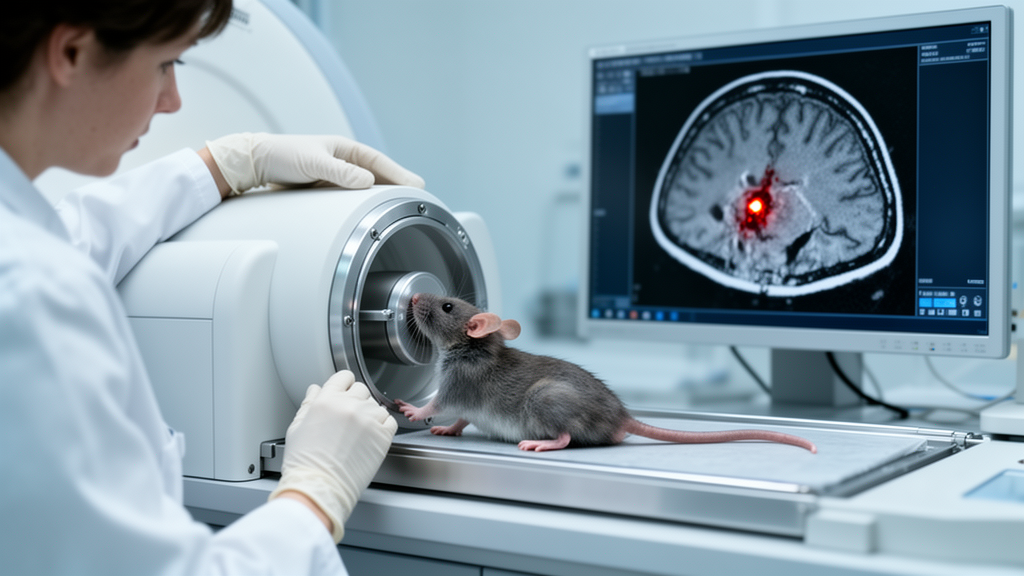
After an ischemic stroke, some damaged brain vessels leak. These tiny bleeds, known as hemorrhagic transformation and microhemorrhages, are scattered through tissue in ways that conventional histology and standard imaging struggle to capture in three dimensions. A new study in Acta Neuropathologica Communications reports a way to see and count them all: ex vivo micro-computed tomography, enhanced with phosphotungstic acid, applied to whole mouse brains after experimental stroke.
The researchers scanned 121 mouse brains collected 1, 3, 7 and 14 days after transient middle cerebral artery occlusion, a standard model of focal cerebral ischemia in which blood flow to a defined brain region is briefly cut off and then restored. Because the technique preserves the entire brain intact, the team could quantify infarction, hemispheric volume changes, hemorrhagic transformation and individual microhemorrhagic lesions within the same three-dimensional anatomical context, rather than inferring whole-organ pathology from thin two-dimensional sections.
The method’s validity rests on careful cross-checks. Hypodense regions on the CT scans, indicating tissue loss, corresponded histologically to areas of neuronal death surrounded by glial scar. Hyperdense regions, which absorb more X-rays, matched clusters of Ter119-positive erythrocytes, confirming that the bright spots were indeed red blood cells and therefore hemorrhages. As an additional test of sensitivity, micro-CT detected aggravated brain edema in Ndrg2 knockout mice, a genetic model of increased swelling, showing the technique can register subtle volume changes as well as overt bleeds.
The scans also captured clinically relevant physiology. Hemorrhagic transformation incidence increased with longer ischemia duration, and mice that developed hemorrhagic transformation showed greater edema and performed worse on rotarod and grid-walk tests of sensorimotor function, linking the imaging findings to measurable behavioral deficits.
The central result, however, is spatial. By analyzing each microhemorrhagic lesion as a discrete three-dimensional object, the researchers uncovered a region-specific temporal pattern during the first week after ischemia-reperfusion: cortical microhemorrhagic burden declined over time, while striatal burden persisted. By day 7, the caudate putamen carried a greater microhemorrhagic load than the cortex.
That regional divergence came with a telling asymmetry in correlations. In the caudate putamen, the number of microhemorrhages correlated positively with both infarct volume and neurological deficit score, suggesting that bleeding in this deep structure tracks the overall severity of injury. In the cortex, no significant correlations were observed, indicating that cortical microbleeds follow a partly independent course.
The authors frame these findings as evidence of spatial heterogeneity in post-ischemic microhemorrhagic burden, a conclusion that was difficult to reach with earlier approaches. Conventional methods generally cannot combine non-destructive preservation of whole-brain three-dimensional anatomy with object-based counting of individual hemorrhagic lesions alongside infarction and edema in the same brain. Contrast-enhanced micro-CT, by contrast, delivers all of these measurements from a single specimen.
For stroke research, the practical implications are substantial. According to the authors, conventional histological and imaging approaches do not readily combine non-destructive preservation of whole-brain three-dimensional anatomical continuity with object-based quantification of individual hemorrhagic lesions alongside infarction and edema in the same experimental brain. A whole-brain, lesion-by-lesion platform makes it possible to ask where bleeds accumulate, how their distribution evolves over days, and how that distribution relates to tissue loss, swelling and functional outcome.
Several caveats deserve emphasis. This is a mouse model of transient focal ischemia, and the measurements were made ex vivo on fixed brains at defined time points rather than in living patients. The behavioral associations come from rotarod and grid-walk performance in the same animals, not from human clinical data. The published version is an accepted-manuscript share, subject to further edits before the final Version of Record.
Even so, the study offers a quantitative template for an underexplored question: not simply whether microbleeds occur after ischemic stroke, but where they occur, how long they persist, and which regional burdens matter most for recovery.
Subject of Research: Three-dimensional quantification of post-ischemic microhemorrhages, edema and infarction in mouse brains using contrast-enhanced micro-CT
Article Title: Three-dimensional quantification using contrast-enhanced micro-CT reveals regional heterogeneity of microhemorrhagic burden after focal cerebral ischemia in mice
Article References: Yang, R., Takarada-Iemata, M., Ishii, H., Wang, M., Fan, Q., Hattori, T., & Hori, O. (2026). Three-dimensional quantification using contrast-enhanced micro-CT reveals regional heterogeneity of microhemorrhagic burden after focal cerebral ischemia in mice. Acta Neuropathologica Communications. https://doi.org/10.1186/s40478-026-02459-9
Image Credits: AI Generated
DOI: 10.1186/s40478-026-02459-9
Keywords: micro-CT, ischemic stroke, microhemorrhage, hemorrhagic transformation, cerebral ischemia, caudate putamen, edema, mouse model, neurovascular, 3D imaging, Three-dimensional, quantification
Cite Scienmag News
Cassandra Pierce. (October 30, 2026). Micro-CT Maps Hidden Brain Bleeds After Stroke in Mice. Scienmag. https://scienmag.com/micro-ct-maps-hidden-brain-bleeds-after-stroke-in-mice/
Cassandra Pierce. "Micro-CT Maps Hidden Brain Bleeds After Stroke in Mice." Scienmag, 30 October 2026, https://scienmag.com/micro-ct-maps-hidden-brain-bleeds-after-stroke-in-mice/. Accessed 1 November 2026.
Cassandra Pierce. "Micro-CT Maps Hidden Brain Bleeds After Stroke in Mice." Scienmag. October 30, 2026. https://scienmag.com/micro-ct-maps-hidden-brain-bleeds-after-stroke-in-mice/