


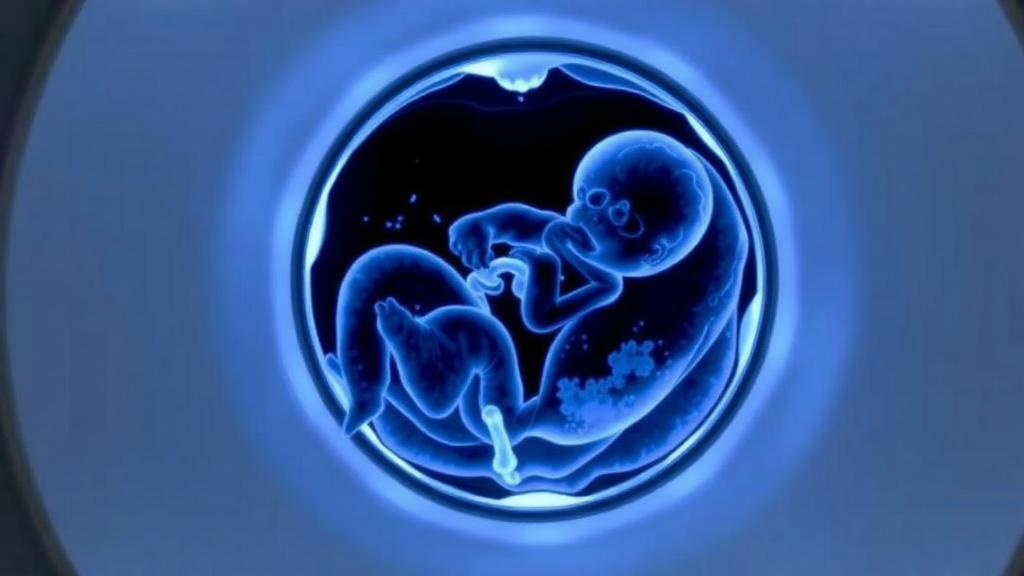
In a striking demonstration of how far molecular imaging has travelled, a team at Copenhagen University Hospital Rigshospitalet has published dynamic PET/CT images of [18F]FDG uptake in a human fetus, using a long-axial field-of-view (LAFOV) scanner. The report, published as an Image of the Month in the European Journal of Nuclear Medicine and Molecular Imaging by Karine Madsen, Kirsten Korsholm, Flemming Littrup Andersen and Thomas Lund Andersen, offers one of the most intimate technological glimpses yet of fetal metabolism in vivo, and it arrives at a moment when next-generation PET hardware is quietly rewriting what clinical nuclear medicine considers possible.
Positron emission tomography depends on a simple but elegant physical principle. A biologically active molecule, in this case fluorodeoxyglucose labelled with the positron-emitting isotope fluorine-18, is injected into the bloodstream. As the radiotracer decays, each positron annihilates with a nearby electron, producing a pair of gamma photons that fly off in nearly opposite directions. Ring-shaped detectors surrounding the patient register these coincident photon pairs, and reconstruction algorithms convert millions of such events into a three-dimensional map of where the glucose analogue has accumulated. Because cells with high metabolic demand, such as tumour cells or inflamed tissue, consume more glucose, FDG PET has become a cornerstone of oncology, cardiology and neurology.
Conventional PET scanners, however, cover only a modest axial length of the body, typically around 15 to 30 centimetres, which means that either the field of view must be moved along the patient or the region of interest must be chosen in advance. LAFOV systems change that equation dramatically. With detector rings extending across much of the body, and with markedly higher sensitivity thanks to a larger solid angle of coverage, these scanners can capture whole-body distributions in a single position, at lower injected activities, in shorter acquisition times, and, crucially, with the temporal resolution needed for true dynamic imaging. It is precisely this combination of sensitivity and continuous coverage that made the Copenhagen team’s fetal study feasible.
Dynamic PET is a fundamentally different exercise from the static snapshots that dominate routine practice. Rather than acquiring a single image after the tracer has distributed, dynamic protocols record the tracer’s journey continuously from injection onward: the bolus arriving through the maternal circulation, the transit across the placenta, and the gradual accumulation and clearance within fetal tissues. From these time-activity curves, quantitative parameters such as uptake rates and glucose metabolism can in principle be derived, rather than merely inferred from a single late time point. In the fetus, where every organ is growing and maturing at a different pace, such kinetic information has long been a tantalising but largely inaccessible prize.
The obstacles to fetal PET are formidable, which is why published examples remain rare. The fetus is a small, moving target suspended in amniotic fluid inside a body that is itself undergoing physiological change. Maternal organs, the placenta and the myometrium all lie in the imaging path, and the fetal signal must be separated from this crowded background. Fetal position shifts and maternal breathing add motion that standard reconstruction pipelines were never designed to handle. Radiation exposure, while modest with modern protocols, must be justified with particular rigour, and the physical dimensions of the pregnant abdomen challenge the geometric assumptions of scanner design. For all these reasons, fetal PET has historically been confined to isolated case reports, usually performed when a clinical question could not be answered by ultrasound or MRI.
That is precisely the context in which the Rigshospitalet report sits. As an Image of the Month contribution, the article documents a single, carefully justified clinical scenario in which the diagnostic value of dynamic FDG PET/CT outweighed the considerations that normally preclude it. The authors obtained written informed consent that explicitly covered the extended scan duration required for research purposes, as well as separate consent for the publication of data and images, underscoring the ethical scaffolding that surrounds any imaging of a pregnant patient. The study received no external funding, and the authors declared that the work itself was free of commercial involvement, although three of the authors noted honoraria from Siemens Healthineers for unrelated academic presentations and, in one case, an ongoing research agreement with the company.
The technical achievement deserves unpacking for readers unfamiliar with the machinery involved. A LAFOV PET/CT scanner pairs the extended PET field of view with computed tomography for anatomical localisation and attenuation correction. The CT component maps how strongly different tissues absorb photons, allowing the reconstruction algorithm to correct for the fact that gamma rays leaving the deep pelvis must traverse more tissue than those leaving superficial structures. In a pregnant patient this correction is especially delicate, because the attenuation landscape changes with fetal and placental position. The high sensitivity of the long-axis design means the injected activity can be kept as low as reasonably achievable, in line with the ALARA principle that governs all radiation medicine, while still collecting enough coincidence events to reconstruct images of diagnostic and scientific quality.
What can dynamic fetal FDG imaging actually reveal? Glucose is the fundamental fuel of the developing brain, and patterns of cerebral FDG uptake reflect the metabolic maturation of neural tissue. The placenta, an organ with its own distinctive glucose handling, appears in the images as a structure whose tracer kinetics can be followed over time. Fetal liver, heart and other organs each leave their metabolic signature. In principle, dynamic acquisition allows clinicians and researchers to distinguish between altered perfusion, altered transport and altered intracellular metabolism, distinctions that static imaging cannot make. For fetal medicine, where conditions such as growth restriction, congenital tumours and metabolic disorders are currently assessed largely through indirect ultrasound markers and MRI morphology, a direct quantitative window onto fetal metabolism represents a genuinely new class of information.
The publication also illustrates a broader shift in the culture of nuclear medicine. As total-body and long-axis PET installations proliferate in academic centres, investigators are revisiting patient populations and physiological questions that were previously off-limits, from ultra-low-dose paediatric studies to kinetic imaging of diffuse disease. The Danish team’s work is a case study in how new hardware creates new scientific opportunities almost as a side effect of its clinical deployment: a scanner bought for oncology becomes, with careful ethics approval and protocol design, an instrument for observing human development. The authors’ data availability statement, which offers access to the datasets on reasonable request, reflects the growing expectation that such rare and valuable imaging records should be shared with the wider research community.
Caution is, of course, the appropriate frame for a single-case Image of the Month. One examination cannot establish reference values, define normal fetal uptake patterns or change clinical guidelines, and the authors themselves present the work as a documentation of feasibility rather than a new standard of care. Yet the historical pattern in medical imaging is that today’s carefully documented case report becomes tomorrow’s protocol. Ultrasound, fetal MRI and even fetal echocardiography all began with isolated demonstrations that the previously invisible could be seen. If LAFOV dynamic PET follows the same trajectory, the Copenhagen images may be remembered as an early, technically demanding first look at the fetal body in motion, organ by organ, molecule by molecule, at the level of its glucose metabolism, a view that no previous generation of imaging technology could provide.
Subject of Research: Dynamic long-axial field-of-view PET/CT imaging of fetal fluorodeoxyglucose uptake
Article Title: Dynamic LAFOV PET/CT imaging of [18F]FDG uptake in the human fetus
Article References: Madsen, K., Korsholm, K., Andersen, F. L., & Andersen, T. L. (2026). Dynamic LAFOV PET/CT imaging of [18F]FDG uptake in the human fetus. European Journal of Nuclear Medicine and Molecular Imaging. https://doi.org/10.1007/s00259-026-08192-8
Image Credits: AI Generated
DOI: 10.1007/s00259-026-08192-8
Keywords: PET/CT, LAFOV PET, FDG, fetal imaging, dynamic PET, molecular imaging, nuclear medicine, fetal metabolism, radiotracer, placenta, prenatal diagnostics, Copenhagen University Hospital
Cite Scienmag News
Ophelia Keating. (September 30, 2026). Long-Axis PET Scanner Captures Rare Dynamic Views of Glucose Uptake in the Human Fetus. Scienmag. https://scienmag.com/long-axis-pet-scanner-captures-rare-dynamic-views-of-glucose-uptake-in-the-human-fetus/
Ophelia Keating. "Long-Axis PET Scanner Captures Rare Dynamic Views of Glucose Uptake in the Human Fetus." Scienmag, 30 September 2026, https://scienmag.com/long-axis-pet-scanner-captures-rare-dynamic-views-of-glucose-uptake-in-the-human-fetus/. Accessed 4 October 2026.
Ophelia Keating. "Long-Axis PET Scanner Captures Rare Dynamic Views of Glucose Uptake in the Human Fetus." Scienmag. September 30, 2026. https://scienmag.com/long-axis-pet-scanner-captures-rare-dynamic-views-of-glucose-uptake-in-the-human-fetus/


