


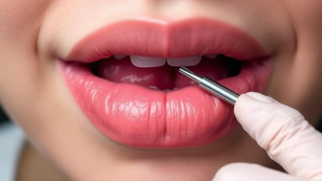
For patients whose nervous systems come under attack before their glands ever complain, one of the most elusive diagnoses in rheumatology may finally have a sharper tool. A new retrospective study from Xuanwu Hospital of Capital Medical University in Beijing suggests that the humble labial gland biopsy, a small incision inside the lower lip to harvest minor salivary glands, carries far more diagnostic weight in suspected Neuro-Sjögren than clinicians have generally assumed. The research, published in Immunity, Inflammation and Disease, analyzed 412 patients evaluated for suspected Sjögren’s disease between January 2019 and December 2023 and found that those with neurological symptoms needed the biopsy more often, and tested positive on it more often, than patients whose disease did not touch the nervous system.
Sjögren’s disease is best known as a chronic autoimmune disorder that dries out the mouth and eyes by destroying exocrine glands, but in a substantial minority of patients it turns its inflammatory sights on the nerves themselves. Neurological involvement is reported in roughly 8 to 49 percent of cases, averaging around 20 percent, and it can strike both the peripheral nervous system, causing neuropathies, and the central nervous system, producing encephalitis, myelitis, or vasculitis. When it does, the consequences can be severe: disability, poor prognosis, and markedly reduced quality of life. Neurologists and rheumatologists call this presentation Neuro-Sjögren, and it is notoriously hard to pin down.
The difficulty lies in the disease’s habit of hiding. Some patients arrive with neurological symptoms as their first and only complaint, lacking the characteristic sicca symptoms of dryness and testing negative for anti-SSA, the antibody that anchors most Sjögren’s diagnoses. Studies cited by the authors indicate that the average diagnostic delay for Neuro-Sjögren stretches from three to six years, and that the wait is even longer in patients who are anti-SSA positive or who first present with neurological symptoms. Every year of delay is a year in which an autoimmune process may be quietly damaging nerves that cannot easily repair themselves.
Enter the labial gland biopsy. The procedure involves removing a small cluster of minor salivary glands from the inner lip and having a pathologist count lymphocytic foci, dense aggregates of at least 50 mononuclear immune cells, per 4 square millimeters of tissue. A focus score of 1 or higher is not diagnostic on its own, but it is a key component of the 2016 ACR/EULAR classification criteria for Sjögren’s disease and can support a clinical diagnosis when combined with other findings. The test is not trivial: it carries risks of local scarring, infection, and permanent numbness, and it demands both an experienced clinician and expert pathological interpretation. That is precisely why knowing who truly benefits from it matters.
The Beijing team, led by Zhen Tian and colleagues, enrolled 456 hospitalized patients with suspected Sjögren’s disease, of whom 412 met the inclusion criteria. Within this cohort, 135 patients had suspected Neuro-Sjögren, meaning their neurological symptoms had been confirmed by a neurologist and, wherever possible, corroborated by objective evidence such as MRI, PET, nerve conduction studies, electromyography, or cerebrospinal fluid analysis. The remaining 277 patients, suspected of Sjögren’s without neurological involvement, served as a comparison group. The researchers deliberately excluded patients whose neurological complaints could be explained by alternative diagnoses, and they excluded anyone with other connective tissue diseases, recent malignancy, active infection, or conditions making biopsy unsafe.
The diagnostic pathway followed real-world practice. Patients who were anti-SSA positive and had objective evidence of glandular impairment could be classified under the 2016 ACR/EULAR criteria without a biopsy. Everyone else, whose serological and glandular findings were insufficient, underwent labial gland biopsy as part of the work-up. Among the 135 suspected Neuro-Sjögren patients, 91 fell into this biopsy-required category, compared with 44 who could be classified without histology. The numbers tell a striking story: biopsy was required in 67.4 percent of suspected Neuro-Sjögren patients versus 56.3 percent of the non-neurological group, a statistically significant difference. Even more compelling, when a biopsy was performed, it came back positive, with a focus score of at least 1, in 81.3 percent of the Neuro-Sjögren group versus just 51.3 percent of the others.
The most eye-catching figure concerns the patients clinicians worry about most. Among anti-SSA-negative patients with suspected Neuro-Sjögren, the biopsy positivity rate reached 77.3 percent. In other words, for roughly three out of four patients who lacked the classic antibody and whose glandular tests were uninformative, the lip biopsy still revealed the focal lymphocytic inflammation that defines Sjögren’s disease. Only 12 patients in the cohort were both anti-SSA negative and biopsy negative, and these individuals could not be classified as having Sjögren’s disease under the 2016 criteria. For a population where diagnosis often stalls for years, that is a meaningful yield from a procedure taking minutes.
Who, then, ends up needing the biopsy? Multivariable logistic regression identified two independent predictors of biopsy requirement: younger age at onset, with an odds ratio of 0.960 per year of age, and early-onset neurological symptoms, meaning neurological manifestations that appeared before the typical sicca symptoms, which more than tripled the odds at an odds ratio of 3.634. In the biopsy-required group, 65.9 percent of patients had presented with neurological symptoms first, compared with 36.4 percent of those classified without biopsy. Conversely, anti-SSB positivity and antinuclear antibody titers of 1:320 or higher were independently associated with a lower likelihood of needing a biopsy, presumably because such patients already carried stronger serological evidence. Elevated IgG showed only a borderline association. The authors are careful to note that these factors reflect their center’s diagnostic pathway, not intrinsic properties of the disease, and should not be read as universal indications for or against biopsy.
Among the patients who actually underwent biopsy, a second pattern emerged. Those with positive results more frequently showed higher erythrocyte sedimentation rates, elevated IgG, and ANA titers of at least 1:320 than biopsy-negative patients. The researchers interpret these as non-specific markers of broader immune activation rather than disease-specific signatures, cautioning that they should never be used in isolation to justify the procedure. Interestingly, while the difference was not statistically significant, biopsy positivity was numerically higher in patients with peripheral nervous system involvement, 87.2 percent, than in those with central nervous system involvement, 77.1 percent, hinting that peripheral phenotypes may be more tightly linked to histopathological glandular inflammation. The study also noted, contrary to much prior literature, a slightly higher proportion of central than peripheral involvement in their cohort, which the authors attribute to center-specific referral patterns and neurological expertise.
The findings align with a growing body of work. Previous studies have shown that positive labial gland biopsies associate with autonomic dysfunction, that more than 90 percent of patients with sensory ganglionopathy test positive on both Schirmer’s test and biopsy despite few sicca symptoms, and that among patients with small fiber neuropathy, up to 30 percent are ultimately diagnosed with Sjögren’s disease. The Beijing study adds a quantitative framework: in serologically ambiguous patients with neurological presentations, the biopsy is not a last resort but frequently the decisive test. The authors acknowledge the limitations of their retrospective, single-center design, including possible selection bias toward more complex inpatient cases and the modest number of outcome events in their regression model. They call for prospective, multicenter studies with richer histological profiling. Still, the practical message for clinicians is clear: when a patient, especially a younger one, presents with neurological symptoms that precede any dryness, and the antibody panel comes back empty, the small glands of the lower lip may hold the answer that blood tests cannot.
Subject of Research: Clinical utility of labial gland biopsy in diagnosing suspected Neuro-Sjögren
Article Title: The Clinical Utility of Labial Gland Biopsy in Patients With Suspected Neuro‐Sjögren
Article References: Tian, Z., Li, X., Li, X., Wang, Y., Wang, C., & Zhao, Y. (2026). The Clinical Utility of Labial Gland Biopsy in Patients With Suspected Neuro‐Sjögren. Immunity, Inflammation and Disease, 14(9), Article e70507. https://doi.org/10.1002/iid3.70507
Image Credits: AI Generated
DOI: 10.1002/iid3.70507
Keywords: Neuro-Sjögren, Sjögren's disease, labial gland biopsy, focus score, anti-SSA, autoimmune disease, peripheral neuropathy, central nervous system, ACR/EULAR criteria, diagnostic delay, small fiber neuropathy, rheumatology
Cite Scienmag News
Ophelia Keating. (October 1, 2026). Lip Biopsy Emerges as a Decisive Clue in the Hidden Nerve Disease of Sjögren’s. Scienmag. https://scienmag.com/lip-biopsy-emerges-as-a-decisive-clue-in-the-hidden-nerve-disease-of-sjogrens/
Ophelia Keating. "Lip Biopsy Emerges as a Decisive Clue in the Hidden Nerve Disease of Sjögren’s." Scienmag, 1 October 2026, https://scienmag.com/lip-biopsy-emerges-as-a-decisive-clue-in-the-hidden-nerve-disease-of-sjogrens/. Accessed 4 October 2026.
Ophelia Keating. "Lip Biopsy Emerges as a Decisive Clue in the Hidden Nerve Disease of Sjögren’s." Scienmag. October 1, 2026. https://scienmag.com/lip-biopsy-emerges-as-a-decisive-clue-in-the-hidden-nerve-disease-of-sjogrens/


