


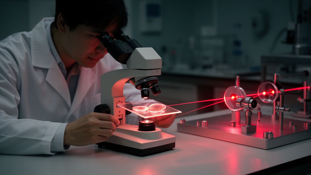
Optical methods have quietly become one of the most productive meeting points between physics and biology. With nothing more exotic than carefully shaped laser light and sensitive detectors, researchers can now read out the orientation of a single collagen fibril, the redox state of an individual mitochondrion, or the vibrational fingerprint of a virus particle — often without staining, fixing, or otherwise disturbing the sample under study. That combination of chemical specificity and physical non-invasiveness is rare in the life sciences, and it explains why biophotonics has moved from a specialist niche to a central tool in everything from fundamental cell biology to clinical diagnostics. A recent Research Topic published in Frontiers in Photonics, titled Advances in Optical Methods for Biological Sample Characterization, gathers four contributions that illustrate both how mature the field has become and how much interpretive and instrumental work still lies ahead.
The collection was curated by Eden Morales-Narváez and Remy Avila of the Centro de Física Aplicada y Tecnología Avanzada at the Universidad Nacional Autónoma de México in Querétaro, together with Pablo Loza-Alvarez of ICFO – Institut de Ciències Fotòniques in Barcelona. When they opened the Research Topic, the editors deliberately set a wide scope, inviting work on bioimaging, biosensors, optical tweezers, particle tracking, and AI-assisted data analysis. In their own Perspective contribution, Avila and colleagues used those five domains as a scaffold for a broader argument: the next substantial gains in optical biological characterization are likely to come less from any single modality reaching a new performance ceiling, and more from combining modalities, from tighter integration with computation, and from a sharper distinction between what an optical signal actually shows and what it is generally assumed to mean. The three remaining contributions each speak to that argument from a different angle, and read together they form a kind of state-of-the-field report with unusually concrete examples.
The first of those examples tackles a problem that has limited the clinical translation of second harmonic generation microscopy for years. SHG is a nonlinear optical process in which certain highly ordered, non-centrosymmetric biological structures — collagen above all — convert two incoming photons into a single outgoing photon of exactly twice the frequency, effectively turning the extracellular matrix into its own microscopic light source. No dye is required, which makes SHG uniquely attractive for imaging living tissue. But there is a catch: in thick tissue, most of the signal propagates forward, in the direction of the illuminating beam, while the only geometry realistically available for in vivo imaging is backward detection, where the signal is collected on the same side the light enters. Backward-detected SHG has historically been harder to interpret than the cleaner forward signal obtained from thin, ex vivo sections, and that interpretive gap has slowed the technique’s move from the bench to the clinic.
To close that gap, Boutkhil and colleagues imaged intact porcine corneas inside a custom pressure-controlled chamber, allowing them to vary intraocular pressure while watching the backward SHG signal change in real time. Their results overturn a long-standing reading of corneal SHG images. The periodic high- and low-intensity bands that had been interpreted in backward images as discrete collagen bundles turn out to be an artifact of collagen crimp — the natural waviness of corneal lamellae. Raise the intraocular pressure and the fibers straighten; the bands fade in step. The banding pattern, in other words, is not a map of anatomical structures but a mechanical signature of how much tension the tissue is under. The authors are candid that a complete quantitative model linking the forward-to-backward signal ratio to tissue structure remains out of reach, but the structural mechanism they have identified should change how the next generation of corneal SHG images — and the diagnostic claims built on them — are read. For a technique whose most promising clinical application is detecting early corneal weakening, that correction matters.
A second axis of the same general problem, quite literally whether to label a sample at all, runs through the review by Cuevas-Galindo and Jiménez-Sánchez. Small-molecule fluorescent probes are among the most indispensable tools in modern biology, but the authors build a sustained case that the very properties that make a probe bright and selective — high-affinity binding, lipophilic partitioning, excited-state reactivity — also make it an active participant in the biology it is meant to report on. The review’s most concrete anchor is its discussion of calcium indicators. These genetically encoded or synthetic dyes work by binding calcium ions, which means they act as buffers inside the cell; their buffering capacity measurably reshapes the very signalling kinetics they are used to track. The measurement instrument, in effect, becomes part of the experiment, and the recorded dynamics can differ from the unperturbed ones in ways that are easy to overlook and hard to correct.
Importantly, the review does not call for abandoning labels. Instead, it positions label-free approaches — autofluorescence imaging of the metabolic coenzymes NAD(P)H and flavins, along with fluorescence lifetime imaging, which exploits the fact that a fluorophore’s decay time is often more informative than its intensity — as a benchmark against which any probe’s effect on underlying physiology should be checked. It also surveys a spectrum of middle-ground strategies designed to narrow the gap between measurement and perturbation. HaloTag and SNAP-tag constructs allow specific proteins to be covalently labelled with tunable dyes; bioorthogonal fluorogenic chemistry keeps probes dark until they react at their cellular target; ratiometric sensing builds an internal reference into the measurement itself. Each strategy represents an engineering compromise between brightness, specificity, and biological neutrality, and the review’s contribution is to make those trade-offs explicit rather than implicit.
The fourth contribution moves from imaging to sensing, and it may be the most immediately striking of the four. Guilcapi and colleagues describe a plasmonic metasurface — a carefully engineered nanostructured surface patterned with Y-shaped nanocavity arrays and tuned to 785-nanometer excitation — that supports surface-enhanced Raman spectroscopy sensitive enough to discriminate human from murine norovirus particles by their vibrational signature alone. SERS works by concentrating electromagnetic fields into nanoscale gaps and cavities, amplifying the intrinsically weak Raman scattering of molecules adsorbed at the surface by many orders of magnitude. Because every biological particle has a characteristic set of vibrational modes, the resulting spectrum functions as a molecular fingerprint. Owing to the structural similarities between the two virus species, this result carries implications well beyond a simple proof of concept: principal component analysis of the SERS spectra recovered two components accounting for over 98 percent of the total variance, enough to separate the species reliably with no antibodies, no PCR, and no labelling step of any kind.
That last point deserves emphasis. Conventional virus identification depends on immunological reagents that bind specific antigens, or on nucleic-acid amplification that requires sample preparation, reagents, and laboratory infrastructure. A label-free plasmonic platform that distinguishes closely related pathogens directly from their spectra could therefore serve as a rapid screening complement in settings where those resources are scarce or turnaround times are critical. Given how disruptive norovirus outbreaks remain for food safety and clinical care — the virus is a recurring cause of gastroenteritis outbreaks in hospitals, cruise ships, and food supply chains — a fast, reagent-free screening route is an appealing prospect. The authors also argue that the underlying nanofabrication approach should generalize to other closely related pathogens, suggesting a platform rather than a single-purpose device.
Taken together, the four contributions converge on a theme that recurs across biophotonics more broadly: an optical signal is rarely as simple to interpret as it first appears. Sometimes the geometry of signal collection changes what a band or a bundle actually represents, as the corneal SHG study demonstrates. Sometimes the probe used to generate contrast measurably perturbs the system it is meant to probe, as the fluorescent labelling review insists. And sometimes telling two nearly identical biological targets apart requires statistical tools as much as photons, as the norovirus biosensor shows. None of the four pieces resolves those tensions outright, but each narrows one of them — by correcting a long-standing source of misreading, by naming a bias that is easy to overlook, by showing that closely related samples can be distinguished without labels, and by setting out a framework against which the field’s broader progress can be measured.
For the wider community, the message is that the frontier of optical biological characterization is as much interpretive as it is instrumental. Better lasers, faster cameras, and smarter algorithms will continue to arrive, but the studies collected here suggest that the field’s most valuable currency in the coming years will be a rigorous understanding of what each photon-based signal genuinely encodes about the underlying biology — and a willingness to question the assumptions baked into decades-old imaging conventions. As the editors close their Research Topic with thanks to the contributing authors and reviewers, the collection stands as a useful reference point for anyone working at the interface of optics and biology, and a reminder that in biophotonics, seeing clearly and interpreting correctly are two separate, equally demanding achievements.
Subject of Research: Advances in optical methods for biological sample characterization, including corneal second harmonic generation imaging, fluorescent labelling trade-offs, and plasmonic SERS virus detection.
Article Title: Editorial: Advances in optical methods for biological sample characterization
Article References: Morales-Narváez, E., Avila, R., & Loza-Alvarez, P. (2026). Editorial: Advances in optical methods for biological sample characterization. Frontiers in Photonics, 7, Article 1917795. https://doi.org/10.3389/fphot.2026.1917795
Image Credits: AI Generated
DOI: 10.3389/fphot.2026.1917795
Keywords: biophotonics, second harmonic generation, corneal imaging, fluorescent probes, label-free detection, autofluorescence, fluorescence lifetime imaging, surface-enhanced Raman spectroscopy, plasmonic metasurface, norovirus, biosensors, bioimaging
Cite Scienmag News
Katie Riggs. (October 15, 2026). Label-Free Light: How Optical Physics Is Rewriting the Rules of Reading Living Tissue. Scienmag. https://scienmag.com/label-free-light-how-optical-physics-is-rewriting-the-rules-of-reading-living-tissue/
Katie Riggs. "Label-Free Light: How Optical Physics Is Rewriting the Rules of Reading Living Tissue." Scienmag, 15 October 2026, https://scienmag.com/label-free-light-how-optical-physics-is-rewriting-the-rules-of-reading-living-tissue/. Accessed 15 October 2026.
Katie Riggs. "Label-Free Light: How Optical Physics Is Rewriting the Rules of Reading Living Tissue." Scienmag. October 15, 2026. https://scienmag.com/label-free-light-how-optical-physics-is-rewriting-the-rules-of-reading-living-tissue/


