


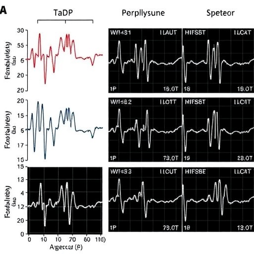
Mitral valve prolapse is often described as a benign heart condition, but a prospective study suggests that a subset of patients may carry a far more serious electrical risk—one that can be exposed by deliberately challenging the heart with carefully timed electrical impulses. In a cohort of 42 people with arrhythmic mitral valve prolapse, nearly half developed sustained ventricular arrhythmias during an invasive electrophysiological test. Those patients were substantially more likely to experience dangerous rhythm disturbances during follow-up, raising the possibility that programmed ventricular stimulation could help doctors identify people at risk of potentially fatal events.
Mitral valve prolapse occurs when one or both flaps of the mitral valve bulge backward into the left atrium as the heart contracts. The condition affects an estimated 0.6 to 2.4 percent of the population and is usually associated with a favorable outlook, particularly when it does not cause severe mitral regurgitation—the backward leakage of blood through the valve—or weaken the left ventricle. Yet some patients develop what specialists call arrhythmic mitral valve prolapse, or AMVP, a phenotype linked to complex ventricular arrhythmias, fainting and, in rare cases, sudden cardiac death.
The distinction matters because the mechanical abnormality of the valve can influence the heart’s electrical behavior even when its pumping function appears relatively preserved. In AMVP, frequent premature ventricular contractions or more complex rhythms such as polymorphic ventricular tachycardia can arise from electrically unstable regions around the mitral valve. A premature ventricular contraction is an early beat originating in the lower chambers rather than following the heart’s normal conduction pathway. When such abnormal beats occur repeatedly or in changing patterns, they can trigger ventricular tachycardia, in which the ventricles beat too rapidly to pump effectively, or ventricular fibrillation, an electrically chaotic rhythm that causes cardiac arrest.
The new study, conducted at University Hospital Münster in Germany and published in Clinical Research in Cardiology, examined 42 consecutive patients who met criteria for AMVP established by the European Heart Rhythm Association. The participants underwent an electrophysiological study between 2016 and 2025. Patients with other structural heart disorders or primary electrical diseases were excluded, allowing the investigators to focus on the relationship between mitral valve prolapse and ventricular rhythm instability. The group had a mean age of 49 years, and about 36 percent were men. Previous fainting had occurred in 40.5 percent of participants, while 69 percent had already shown episodes of nonsustained ventricular tachycardia.
During programmed ventricular stimulation, doctors delivered a sequence of electrical pulses through a catheter positioned in the right ventricle. The stimulation began with regular impulses and then introduced one or two premature beats at progressively shorter intervals. This procedure is designed to probe how readily the heart’s electrical system breaks into a sustained abnormal rhythm. In the study, impulses were delivered from both the right ventricular apex and the right ventricular outflow tract, using baseline cycle lengths ranging from 500 to 330 milliseconds and coupling intervals as short as 200 milliseconds. Patients stopped beta-blockers or antiarrhythmic drugs for at least five half-lives before testing, reducing the chance that medication would suppress inducibility.
The test triggered sustained ventricular arrhythmias in 20 of the 42 patients, or 47.6 percent. Every induced event was either polymorphic ventricular tachycardia or ventricular fibrillation; no monomorphic ventricular tachycardia was observed. The distinction is technically important. Monomorphic ventricular tachycardia typically follows a stable electrical circuit, often around a scar, whereas polymorphic ventricular tachycardia changes its electrical pattern from beat to beat and can deteriorate into ventricular fibrillation. In mitral valve prolapse, the latter rhythms are thought to reflect a more diffuse and dynamic electrical vulnerability rather than a single fixed re-entry pathway.
The patients were then monitored for an average of 38 months, with a wide variation of roughly two years in either direction. Following discussions with their physicians, some received an implantable cardioverter-defibrillator, or ICD, while others received an implantable loop recorder. An ICD continuously monitors the heart and can deliver pacing or a shock when it detects a life-threatening rhythm. A loop recorder is less invasive and records the heart’s electrical activity over long periods, helping physicians capture intermittent events that might otherwise escape routine examinations.
Over follow-up, clinically significant ventricular arrhythmias occurred in nine patients who had developed an arrhythmia during programmed stimulation—45 percent of that group—compared with only two patients, or 9.1 percent, among those whose stimulation test did not induce a pathological rhythm. The difference was statistically significant, with a probability value of 0.01. When the investigators adjusted for other variables in a multivariable logistic regression analysis, inducibility remained associated with subsequent clinically important arrhythmias. The event rate was 19.1 arrhythmias per 100 patient-years after a pathological test, compared with 5.0 per 100 patient-years after a negative test.
The implanted defibrillators delivered 11 appropriate therapies to seven patients. Of those recorded episodes, 36.4 percent were ventricular fibrillation and 63.6 percent were polymorphic ventricular tachycardia. Six ICD recipients with an abnormal stimulation test received 10 of the therapies, while one of eight ICD recipients with a negative test experienced a recurrence. Loop recorders also documented high-risk rhythms more often in patients with abnormal electrophysiological studies. No deaths occurred during the observation period, an encouraging result but one that also means the study cannot establish whether the test predicts mortality specifically.
The researchers also used cardiac magnetic resonance imaging to search for anatomical and tissue features associated with electrical instability. Scans were available for 41 participants. Significant mitral annular disjunction, a separation between the posterior mitral valve leaflet and the adjacent ventricular muscle during contraction, was present in 56.1 percent. Late gadolinium enhancement, a magnetic resonance marker of replacement fibrosis, appeared in 61 percent. Fibrosis around the mitral apparatus and papillary muscles can disrupt the normal spread of electrical impulses, creating regions in which conduction slows or becomes uneven. Mechanical stretching may contribute to this process: when the valve and annulus move abnormally, the inferobasal left ventricle and posterior papillary muscle can experience repeated stress that promotes microscopic injury and scar formation.
Previous fainting was the only baseline feature that significantly correlated with whether an arrhythmia could be induced during testing. That finding fits with clinical experience, because unexplained syncope may be the outward sign of a brief, self-terminating ventricular rhythm. However, the study was not large enough to determine how programmed stimulation compares with each imaging or electrocardiographic marker, or whether combining them would produce a more accurate risk score. Current assessment already considers factors such as bileaflet prolapse, frequent or complex ventricular ectopic beats, T-wave inversions on a 12-lead electrocardiogram, mitral annular disjunction and fibrosis visible on magnetic resonance imaging.
Programmed ventricular stimulation has been used for decades in other cardiac disorders, particularly in patients with previous heart attacks and reduced ventricular function. In those settings, the test is often aimed at provoking monomorphic ventricular tachycardia associated with scar-based re-entry. Its role in AMVP has been uncertain partly because earlier studies were small, retrospective or conducted with stimulation protocols more aggressive than those used in contemporary practice. A 1985 study reported inducibility in 45 percent of selected patients with mitral valve prolapse and ventricular arrhythmias, but often required three premature stimuli. Another study from the 1980s found sustained arrhythmias in 15 of 36 patients, many of whom had already experienced fainting, arrhythmias or sudden cardiac arrest. By contrast, a 2021 study of people with Barlow disease found sustained arrhythmias in only four of 21 patients and did not assess long-term outcomes.
The new findings therefore offer something older studies could not: prospective follow-up linking the result of the invasive test to later clinical events. Still, they do not mean that every person with mitral valve prolapse should undergo catheter-based stimulation or receive an ICD. The cohort was drawn from a single specialist center and included patients already considered to have the arrhythmic phenotype, so the results cannot be applied directly to the far larger population with uncomplicated MVP. Device programming was left to treating physicians, and more aggressive ICD settings may have produced therapies for rhythms that were not necessarily clinically dangerous. In addition, because all induced rhythms were polymorphic tachycardia or fibrillation, the test’s specificity may be lower than it would be for inducible monomorphic ventricular tachycardia.
The findings could nevertheless change how specialists approach patients who fall between clearly low- and high-risk categories. An individual with mitral valve prolapse, fainting, complex ventricular beats and suggestive magnetic resonance findings may not fit neatly into existing recommendations for either immediate ICD implantation or simple observation. In such cases, an abnormal stimulation test might support closer surveillance, an implantable loop recorder or preventive defibrillator therapy, while a negative result could help inform a more conservative strategy—although it cannot guarantee that a dangerous arrhythmia will never occur. The study’s authors emphasize that larger, multicenter investigations are needed before programmed ventricular stimulation can become a routine part of AMVP risk assessment. For now, the test appears less like a universal screening tool than a potentially valuable piece of a highly specialized diagnostic puzzle: a controlled electrical stress test that may reveal which apparently stable hearts are poised to become dangerously unstable.
Cite Scienmag News
Ophelia Keating. (August 26, 2026). Inducible ventricular arrhythmias predict clinical events in mitral valve prolapse syndrome. Scienmag. https://scienmag.com/inducible-ventricular-arrhythmias-predict-clinical-events-in-mitral-valve-prolapse-syndrome/
Ophelia Keating. "Inducible ventricular arrhythmias predict clinical events in mitral valve prolapse syndrome." Scienmag, 26 August 2026, https://scienmag.com/inducible-ventricular-arrhythmias-predict-clinical-events-in-mitral-valve-prolapse-syndrome/. Accessed 7 September 2026.
Ophelia Keating. "Inducible ventricular arrhythmias predict clinical events in mitral valve prolapse syndrome." Scienmag. August 26, 2026. https://scienmag.com/inducible-ventricular-arrhythmias-predict-clinical-events-in-mitral-valve-prolapse-syndrome/


