


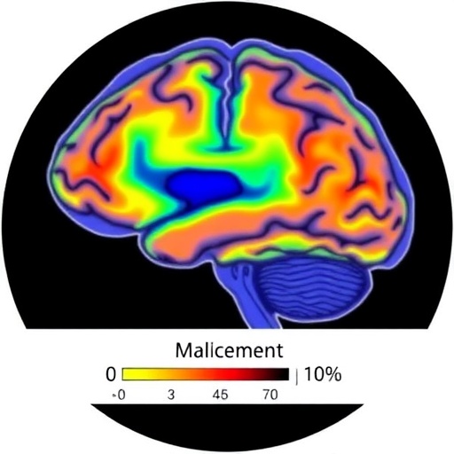
A new study is drawing attention to one of the brain’s most closely watched structures in the search for early warning signs of bipolar disorder. Published in Translational Psychiatry, the research examines how the volumes of specific hippocampal subregions differ in young people considered at high risk for bipolar disorder, both before treatment and after receiving antidepressant medication. The work, led by B. Tang, L. R. Patino and T. J. Blom, places the hippocampus at the center of a question with major clinical implications: can measurable changes in brain anatomy help researchers understand vulnerability to bipolar illness, treatment response and the possible biological effects of antidepressants in young people?
The hippocampus is a curved structure located deep within the brain’s temporal lobes and is best known for its role in forming and retrieving memories. It is not a single, uniform unit. Instead, it contains several interconnected subregions, including the dentate gyrus, CA fields and the subiculum. These areas participate in different aspects of memory, learning, stress regulation and emotional processing. Because bipolar disorder involves disturbances in mood, cognition and stress sensitivity, scientists have increasingly turned to the hippocampus as a potential source of biological clues. Examining the structure as a collection of smaller regions may reveal patterns that disappear when the entire hippocampus is treated as one volume.
The study focuses on youth at high risk for bipolar disorder, a group that can include individuals with a strong family history or early symptoms associated with future mood illness. Being at high risk does not mean that a person will inevitably develop bipolar disorder. Rather, it identifies a population in which researchers can investigate vulnerability before the full disorder emerges. This distinction is crucial. Studying people before repeated mood episodes, long-term medication exposure or years of illness-related stress may offer a clearer view of the biological factors that precede diagnosis.
The research compares hippocampal subregion volumes at baseline and following antidepressant treatment. In practical terms, the investigators are asking whether the anatomy of these finely defined brain regions differs at the beginning of treatment and whether it changes over time. Such comparisons can help separate features that may reflect pre-existing risk from changes that emerge during clinical care. They can also provide insight into whether antidepressant treatment is associated with detectable structural differences, although brain-volume measurements alone cannot establish that a medication caused a particular change.
This question is especially important because antidepressants occupy a complicated place in the treatment of young people vulnerable to bipolar disorder. Antidepressants are commonly used to treat depressive symptoms, but clinicians must carefully monitor patients who may later develop bipolar-spectrum illness. Some individuals can experience mood destabilization, agitation or a shift toward manic symptoms, while others may benefit without such complications. The study does not turn a brain scan into a simple test for deciding who should receive medication. Instead, it contributes to a broader effort to understand how treatment, development and inherited risk interact inside the brain.
Hippocampal volume is also influenced by many factors unrelated to bipolar vulnerability or antidepressant exposure. Age, puberty, sex, sleep, chronic stress, trauma, exercise, substance use and other medications can all affect brain structure or the quality of neuroimaging measurements. The hippocampus continues to mature during adolescence, making timing particularly important when researchers compare young participants across months or years. Advanced imaging can estimate the size of individual subregions, but these measurements remain sensitive to scanning protocols, image resolution and the methods used to define anatomical boundaries.
The significance of the paper therefore lies not in the idea that one subregion can provide a definitive diagnosis, but in the possibility that patterns across several regions may improve scientific understanding of risk. If certain hippocampal differences are consistently observed in high-risk youth, they could point toward biological pathways involved in mood regulation and memory-related symptoms. If volumes change after treatment, those findings could suggest that the brain remains structurally responsive during adolescence and that medication-related effects deserve closer study. Neither result, however, would by itself predict an individual’s future or prove that structural change equals clinical improvement.
The work also highlights a major challenge in psychiatric neuroscience: translating group-level differences into useful information for individual patients. A statistically meaningful difference between groups may be too small or variable to guide decisions for one teenager. Researchers must also determine whether anatomical changes are linked to measurable changes in depression, anxiety, cognition, sleep or emerging manic symptoms. Longitudinal studies, repeated assessments and larger samples will be essential for establishing whether hippocampal measurements remain stable markers of vulnerability, track the course of illness or reflect temporary effects associated with treatment and development.
For families and clinicians, the findings should be viewed as an important research step rather than an immediate clinical tool. Brain imaging currently cannot diagnose bipolar disorder before symptoms appear, and no young person should be labeled on the basis of hippocampal volume alone. The value of studies like this is cumulative: by mapping how vulnerable brains differ and respond over time, researchers may eventually identify more precise biological subtypes, improve monitoring and develop treatments tailored to the earliest stages of mood illness. The hippocampus may not deliver a single dramatic answer, but its subregions could help reveal how risk and treatment are written into the developing brain.
Cite Scienmag News
Glenn Wilkins. (August 1, 2026). Hippocampal Subregion Changes Before and After Antidepressants in Youth at Bipolar Risk. Scienmag. https://scienmag.com/hippocampal-subregion-changes-before-and-after-antidepressants-in-youth-at-bipolar-risk/
Glenn Wilkins. "Hippocampal Subregion Changes Before and After Antidepressants in Youth at Bipolar Risk." Scienmag, 1 August 2026, https://scienmag.com/hippocampal-subregion-changes-before-and-after-antidepressants-in-youth-at-bipolar-risk/. Accessed 3 September 2026.
Glenn Wilkins. "Hippocampal Subregion Changes Before and After Antidepressants in Youth at Bipolar Risk." Scienmag. August 1, 2026. https://scienmag.com/hippocampal-subregion-changes-before-and-after-antidepressants-in-youth-at-bipolar-risk/


