


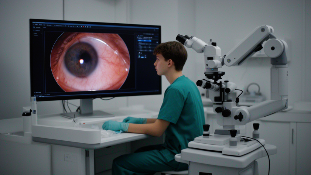
A prospective crossover study has compared how medical students and ophthalmology residents learn microsurgical skills on a virtual reality simulator when the same procedures are displayed through a conventional operating microscope versus a modern 3D heads-up system. The study, published in BMC Medical Education, found that the two visualization technologies produced broadly comparable objective performance, with one notable exception: participants scored significantly better in a cataract surgery module when using the conventional ocular microscope, while results in a vitrectomy module were statistically indistinguishable between the two systems.
The research team, led by Franziska Eckardt, Maximilian-Joachim Gerhardt and Siegfried Priglinger, enrolled 32 participants with no prior microsurgical experience: 16 medical students who had some exposure to ophthalmology and 16 residents. Each participant completed standardized cataract and vitrectomy training modules on the EyeSi surgical simulator, manufactured by Haag-Streit in Köniz, Switzerland, once under the conventional microscope and once with a 3D display. Because every trainee used both systems, the crossover design allowed direct within-participant comparison. The investigators recorded objective performance parameters scored by the simulator and collected subjective evaluations through questionnaires covering safety, handling, comfort, learning progress and interaction with instructors.
The objective results were mixed but largely reassuring for advocates of heads-up technology. In the vitrectomy simulation, scores did not differ significantly between the two visualization systems, with a p-value of 0.126. In the cataract simulation, however, the conventional microscope achieved significantly higher scores, with a p-value of 0.023. Across both modules combined, the overall task scores showed no significant difference between the systems, at p = 0.376.
Where the 3D heads-up system clearly stood out was in subjective ratings. Participants rated it significantly superior in terms of control, with p = 0.013; instrument handling, with p = 0.033; and movement precision, with p < 0.001. It was also rated better for comfort and for interaction with the instructor, both reaching p < 0.001.
Preferences for one system over the other diverged by level of training. Students tended to prefer the 3D system, whereas residents more often preferred the conventional microscope, although this difference in preference was not statistically significant, with p = 0.183. The authors interpret this pattern as a possible habituation effect: residents, having spent more time at the slit lamp and with conventional visualization techniques, may simply be more familiar with the traditional arrangement, while students arrive without those learned habits and adapt readily to the digital display.
Notably, every participant in the study reported that skills acquired while training with the 3D system could be transferred to the conventional microscope. This perceived transferability matters for training programs, because it suggests that introducing trainees to microsurgery through a heads-up display is unlikely to lock them out of conventional operating environments, even if it does not yet match the conventional microscope for objective cataract simulator performance.
The findings arrive as 3D digital visualization systems are becoming increasingly integrated into contemporary ophthalmic surgery. The authors argue that early exposure to heads-up technology during simulator-based training may help familiarize trainees with these modern visualization techniques before they encounter them in the operating room.
The study’s conclusions are bounded by its design and scale. It was a single-center trial with 32 participants, all novices to microsurgical simulation, and its outcome measures were simulator scores and subjective questionnaires rather than performance in real operations. The authors explicitly state that future clinical studies are required to determine whether these simulator-based findings translate into improved operative performance in actual surgery. The published version is an open-access article released early as a citable accepted manuscript, subject to further editorial processing before the final Version of Record appears.
For surgical educators, the study offers a pragmatic message. The 3D heads-up display achieved comparable simulator performance in vitrectomy training and somewhat lower performance in cataract training compared with the conventional microscope, while trainees consistently rated it better for comfort, precision, control and interaction with instructors. Given the varying preferences between students and residents and the possible role of habituation, the results do not declare one technology superior overall, and the authors note only that early simulator exposure to heads-up systems may familiarize trainees with visualization techniques that are increasingly common in contemporary ophthalmic surgery.
Subject of Research: Comparison of conventional operating microscopy and 3D heads-up visualization for teaching ophthalmic microsurgical skills on a virtual reality simulator
Article Title: Teaching ophthalmic microsurgical skills: conventional microscopy versus heads-up visualization
Article References: Eckardt, F., Gerhardt, M. J., & Priglinger, S. (2026). Teaching ophthalmic microsurgical skills: conventional microscopy versus heads-up visualization. BMC Medical Education, 26(1), Article 1403. https://doi.org/10.1186/s12909-026-10269-9
Image Credits: AI Generated
DOI: 10.1186/s12909-026-10269-9
Keywords: ophthalmology, microsurgery, surgical training, 3D heads-up visualization, virtual reality simulator, cataract surgery, vitrectomy, medical education, EyeSi simulator, ergonomics, Teaching, ophthalmic
Cite Scienmag News
Ophelia Keating. (October 23, 2026). Heads-up 3D display matches conventional microscope in simulator eye surgery training. Scienmag. https://scienmag.com/heads-up-3d-display-matches-conventional-microscope-in-simulator-eye-surgery-training/
Ophelia Keating. "Heads-up 3D display matches conventional microscope in simulator eye surgery training." Scienmag, 23 October 2026, https://scienmag.com/heads-up-3d-display-matches-conventional-microscope-in-simulator-eye-surgery-training/. Accessed 23 October 2026.
Ophelia Keating. "Heads-up 3D display matches conventional microscope in simulator eye surgery training." Scienmag. October 23, 2026. https://scienmag.com/heads-up-3d-display-matches-conventional-microscope-in-simulator-eye-surgery-training/


