


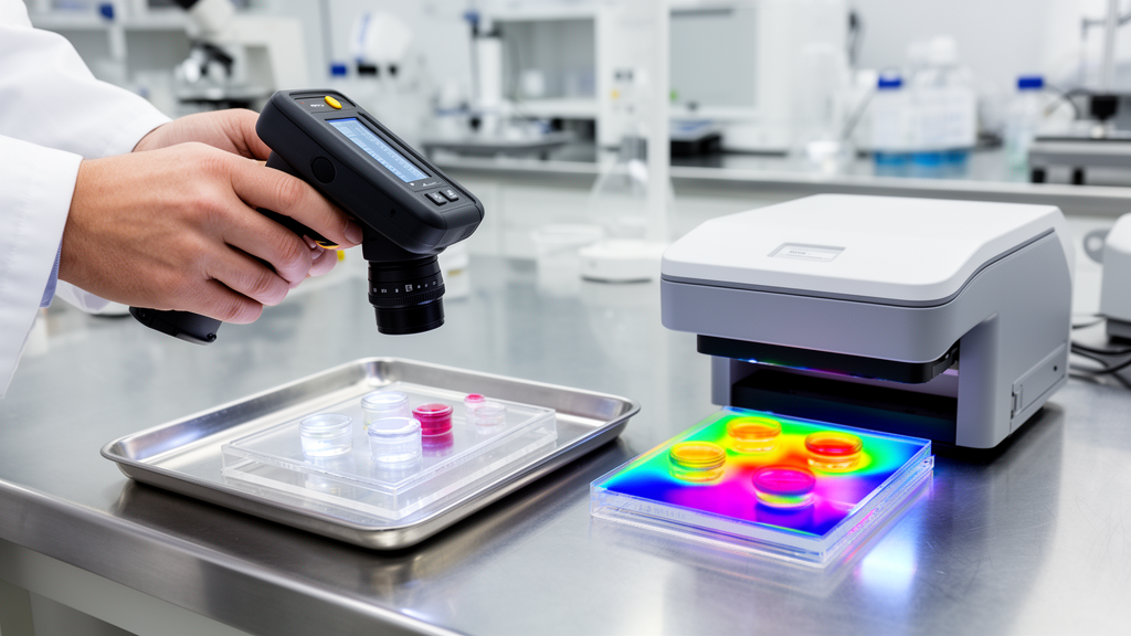
Researchers have developed a handheld mid-infrared imaging spectrometer that produces label-free chemical maps of human tissue and other samples with performance comparable to a much larger laboratory instrument. The experimental system combines visible and mid-infrared laser light in a flexible probe measuring 8 by 8 inches, while a compact base unit houses the lasers and control electronics. In side-by-side tests with a state-of-the-art benchtop photothermal mid-infrared spectroscopic imaging system, the miniaturized device delivered comparable chemical contrast, spectral detail and spatial resolution.
The researchers describe the result as a technical foundation for portable chemical imaging, although the platform remains a research prototype and has not been validated for clinical diagnosis or use during surgery. The design could eventually give surgeons a way to examine freshly removed tissue without waiting for laboratory results, research team leader Rohith Reddy of the University of Houston said in a news release accompanying the peer-reviewed publication in Optica. A proposed clinical application would involve scanning excised tissue to help assess whether it is malignant or whether cancer cells remain at a surgical margin.
That possibility is not a demonstrated clinical outcome: the reported work involved imaging laboratory samples and comparing instruments, not surgery, patient outcomes or diagnostic decisions. Before clinical use, Reddy said the system’s repeatability, safety and diagnostic performance would need thorough evaluation under realistic clinical conditions. The handheld instrument is a compact version of photothermal mid-infrared spectroscopic imaging, or MIRSI, a method that maps the molecular composition of a specimen by measuring localized changes induced by absorbed infrared energy.
Molecules absorb mid-infrared light at wavelengths associated with their chemical bonds, creating characteristic spectra that can distinguish substances such as proteins, lipids and nucleic acids. In photothermal imaging, the instrument detects tiny heat-induced changes rather than forming an image directly from the infrared signal. This approach can provide chemically specific images, but conventional systems are generally bulky, laboratory-based instruments.
The original MIRSI platform occupied more than 9 square feet, compared with the 8-square-inch imaging probe developed in the new study. Miniaturizing the system required the researchers to solve a difficult optical problem. MIRSI uses both visible and mid-infrared illumination, and the two beams must be focused onto the same location.
Materials that transmit visible light effectively often absorb mid-infrared radiation, while optical materials suited to the infrared can introduce dispersion and other wavelength-dependent distortions that degrade the signal. The researchers avoided needing a single lens material that worked across both wavelength ranges by replacing conventional lenses with mirrors. Because mirrors can reflect visible and mid-infrared light, both beams could follow the same optical path and then be directed by an off-axis parabolic mirror onto the specimen.
The system also uses raster scanning so that the focused beams collect data across the sample. To make the probe flexible, the team used chalcogenide optical fibers to carry the mid-infrared light directly from the laser, eliminating bulky free-space optics along that part of the system. Visible light from a red diode laser was combined with light from a modulated quantum cascade laser, and both were delivered through the fiber tether to the handheld imaging head.
The beams were brought together with a short-pass dichroic mirror before reaching the shared focusing optics. Researchers said careful design and alignment were needed to limit image distortions that mirrors can introduce. The team tested the handheld system with human cervical and ovarian cancer tissues, a human bone-marrow biopsy specimen and mouse kidney tissue.
The study was therefore an experimental laboratory evaluation involving human tissue samples and an animal tissue sample, not a clinical trial in people. The researchers also imaged PMMA and polystyrene, two polymers with distinct mid-infrared signatures, as controlled tests of chemical specificity. Each sample was then analyzed with a state-of-the-art benchtop chemical imaging system.
Across these comparisons, the handheld system produced spectra and images with chemical contrast and imaging performance comparable to those obtained by the larger instrument, indicating that miniaturization had preserved the method’s core analytical capabilities on the tested material. The reported spatial resolution was as fine as 2 micrometers, which the researchers said was five times finer than direct infrared detection permits. The handheld system also matched or exceeded current benchtop instruments in spectral and spatial performance.
Quantitative comparisons showed that tissue spectra from the portable system agreed with reference Fourier-transform infrared measurements at a cosine similarity of 0.935. Chemical images produced with the handheld scanner correlated with the benchtop results at r = 0.90. These results support the conclusion that the compact probe can recover chemical information similar to that obtained from the laboratory system on the tested samples; they do not establish that the device can diagnose cancer or perform other clinical tasks accurately in routine use.
One remaining limitation is the instrument’s spectral range, which currently extends from 1,150 to 1,400 inverse centimeters. The researchers are working to broaden that mid-infrared bandwidth so the system can capture more complete molecular signatures and improve its ability to distinguish different biochemical constituents. They also plan to increase imaging speed.
Faster acquisition would reduce motion artifacts and could make genuinely freehand scanning more practical for clinicians and technicians, but the present system has not yet achieved that goal. The study therefore demonstrates technical feasibility rather than a completed surgical tool. The prototype also points toward possible applications beyond medicine, including pharmaceutical quality control, polymer manufacturing and forensic analysis, situations in which chemical composition must be measured outside a specialized laboratory.
Those uses remain potential applications rather than validated deployments. The device requires no stains or labels to create its chemical maps, and the study suggests that this label-free capability can survive miniaturization. Further work must address the restricted spectral bandwidth, acquisition speed, repeatability, safety and diagnostic performance, as well as how the scanner behaves with moving tissue and operators handling it in real-world environments.
If those challenges can be resolved, the compact MIRSI system could offer a portable way to examine molecular makeup closer to where a sample is collected, potentially reducing the interval between examining tissue and obtaining laboratory-style chemical information without implying that the current prototype has already demonstrated clinical benefit.
Subject of Research: Laboratory development and validation of a handheld label-free mid-infrared chemical imaging system for tissue analysis.
Article Title: New handheld device maps chemical composition using infrared light
Article References: New handheld device maps chemical composition using infrared light. Original publication (Bibliographic details unavailable.)
Image Credits: AI Generated
DOI: Not provided
Keywords: handheld imaging, mid-infrared spectroscopy, chemical mapping, label-free tissue imaging, MIRSI, optical engineering, quantum cascade laser, cancer tissue, research prototype, clinical validation, handheld, device
Cite Scienmag News
Nathaniel Bowman. (October 26, 2026). Handheld infrared imager matches laboratory chemical mapping performance. Scienmag. https://scienmag.com/handheld-infrared-imager-matches-laboratory-chemical-mapping-performance/
Nathaniel Bowman. "Handheld infrared imager matches laboratory chemical mapping performance." Scienmag, 26 October 2026, https://scienmag.com/handheld-infrared-imager-matches-laboratory-chemical-mapping-performance/. Accessed 26 October 2026.
Nathaniel Bowman. "Handheld infrared imager matches laboratory chemical mapping performance." Scienmag. October 26, 2026. https://scienmag.com/handheld-infrared-imager-matches-laboratory-chemical-mapping-performance/


