


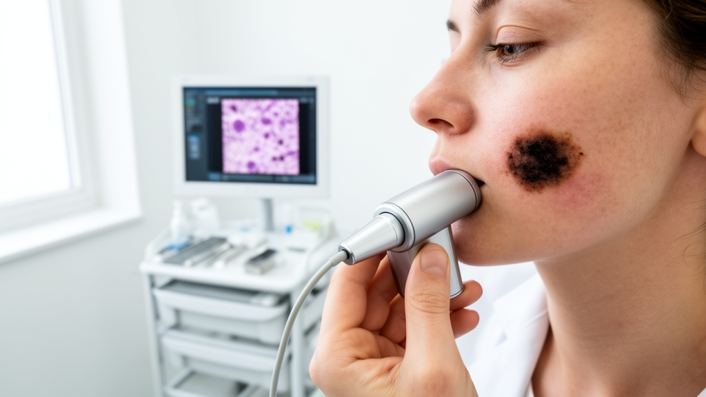
Lentigo maligna, a slow-growing precursor of melanoma that typically appears as an irregular pigmented patch on sun-damaged skin of the face, has long frustrated surgeons for one deceptively simple reason: nobody can see exactly where it ends. The tumor spreads invisibly along the superficial layers of chronically sun-exposed skin, often extending millimeters or even centimeters beyond the brown discoloration that clinicians can detect with the naked eye or a dermatoscope. When the disease comes back after an initial excision, the challenge becomes even harder, because surgical scarring distorts the skin’s architecture and masks the subtle clues dermatologists normally rely on. A new prospective study from the University of Bologna and its affiliated IRCCS hospital, published in the Archives of Dermatological Research, suggests that a handheld imaging device can solve part of this problem, allowing surgeons to trace the tumor’s true footprint before the first incision and dramatically reducing the scale of the operations that follow.
The study, led by Federico Venturi and Emi Dika with colleagues in Bologna and Ancona, focused specifically on recurrent lentigo maligna of the face. Adults who had previously undergone complete surgical excision of a facial lentigo maligna and then developed a histologically confirmed recurrence at the same site within 24 months were enrolled. Recurrence was defined strictly: a new lesion had to arise after a documented disease-free interval and be verified by biopsy or excision. This is a notoriously difficult patient population, because the previous surgery leaves fibrotic tissue that both conceals subclinical tumor strands and complicates any attempt to judge margins by eye. The researchers compared two management pathways in a prospective, single-center design: one group of 21 patients had their surgical margins mapped preoperatively with handheld reflectance confocal microscopy, while a control group of 33 patients was planned using standard clinical and dermoscopic assessment alone.
Reflectance confocal microscopy is a noninvasive optical technique that uses a low-power laser focused on a single point within the skin and measures the light scattered back from tissue structures. By scanning the beam laterally and at different depths, the device builds grayscale horizontal images, sometimes described as optical sections, at near-cellular resolution down to roughly the depth of the superficial dermis. In the hands of a trained reader, this makes it possible to identify the hallmark features of lentigo maligna in vivo: proliferation of atypical, bright, round and dendritic cells along the basal layer of the epidermis, irregular honeycomb or disordered patterns of the epidermis, and pagetoid spread of melanocytes upward through the epidermal layers. Crucially, the handheld version of the device trades some of the stability of traditional benchtop confocal systems for portability, allowing clinicians to examine patients directly in the clinic or at the bedside and to probe skin anywhere on the body, including curved facial contours that a fixed microscope cannot easily reach.
The Bologna team used a standardized adhesive tape technique to translate confocal findings into a practical surgical map. After identifying the outermost confocal extension of the tumor, the clinicians marked the skin and applied adhesive suture tapes bearing grid markings, creating a durable record of the mapped boundary that could be handed directly to the surgeon. This approach, which the group has refined in earlier work on non-melanoma skin cancers of the head and neck, effectively converts a live optical examination into a physical template for excision. In the study, surgical margins were subsequently verified by permanent-section histopathology in every case, meaning the accuracy of the preoperative maps could be judged against the gold standard of laboratory examination of the removed tissue.
The results were striking. Complete margin clearance in a single surgical stage was achieved in 85.7 percent of patients in the confocal-mapping group, compared with 60.6 percent of those planned conventionally. In other words, roughly nine out of ten patients whose tumors had been mapped with the handheld device left the operating room after one procedure, while nearly four in ten of the conventionally planned patients needed additional excisions to reach clear margins. Each repeat excision carries costs beyond the operating schedule: more anesthesia, more scarring on cosmetically sensitive facial skin, longer healing times, and greater patient anxiety. Avoiding staged procedures is therefore one of the most meaningful outcomes a preoperative planning tool can deliver.
The size of the surgical defects told a similar story. The mean defect diameter in the confocal group was 1.7 centimeters, with a standard deviation of 0.6, versus 2.4 centimeters, with a standard deviation of 0.8, in the clinical group, a difference that reached statistical significance at p less than 0.01. Smaller defects matter enormously on the face, where the availability of local tissue governs what kind of closure is possible. This translated directly into the study’s primary outcome: only 28.6 percent of patients in the confocal-mapped group required invasive reconstructive procedures, such as flaps or grafts, compared with 60.6 percent of the conventionally planned group, a reduction that was statistically significant at p equal to 0.009. The authors describe this as downstaging the surgical repair, a phrase that captures the practical benefit: the same tumor can be managed with a simpler, more conservative operation when its boundaries are known in advance.
Perhaps the most important question for any technique that narrows surgical margins is whether it compromises cancer control. On this point the study was reassuring, at least over the short follow-up window. Local recurrence rates at 12 months were low and statistically comparable between the two groups, at 4.7 percent in the confocal group and 6.1 percent in the clinical group. The investigators emphasize that the confocal approach did not cut corners oncologically; rather, it delineated the subclinical extension of the tumor more accurately than visual inspection, so the excision could be tailored to the disease rather than to a generous default margin. Because lentigo maligna can recur years after treatment, longer follow-up will be needed to confirm that the early equivalence holds, and the authors themselves frame the recurrence data as short-term oncologic control.
The findings arrive amid a growing body of evidence supporting confocal margin mapping for this tumor type. Previous prospective work has shown that confocal delineation of lentigo maligna margins correlates well with staged excision histology, and a 2025 systematic review and meta-analysis concluded that reflectance confocal microscopy is a reliable tool for margin assessment in lentigo maligna melanoma. A Dutch group has gone further, reporting the successful implementation of handheld confocal microscopy as a standard of care in the surgical management of lentigo maligna at their head and neck center. The Bologna study adds a specific and clinically valuable data point: the technique works even in the hardest scenario, recurrent disease at a previously operated site, where scarring has traditionally made margin assessment least reliable.
The broader significance of the study lies in what it suggests about the future of skin cancer surgery. Noninvasive imaging modalities, including reflectance confocal microscopy, optical coherence tomography, and dermoscopy-guided high-frequency ultrasound, are increasingly being tested as ways to plan excisions of skin cancers before the patient reaches the operating table. For lentigo maligna in particular, where histologic margins routinely exceed clinical margins by wide margins, the economic and human calculus is compelling: a handheld device examination in clinic is far cheaper and less burdensome than a second operation, a flap reconstruction, or a prolonged staged excision process. The adhesive tape mapping method also lowers the barrier to adoption, since it requires no exotic materials and integrates with existing surgical workflows.
Limitations remain, and the authors are careful about them. The study was single-center, the two groups were not randomized in the traditional sense, and the confocal cohort was smaller than the comparison cohort, all of which counsel caution before declaring a new standard of care. Reader expertise is also a genuine constraint, because interpreting confocal images of sun-damaged facial skin requires training and experience, and false negatives at the margin edge remain a risk. Still, the direction of the evidence is consistent across multiple independent groups, and the Bologna data address the scenario where the clinical need is most acute. For elderly patients with recurrent facial lentigo maligna, many of whom have limited tissue reserve and significant comorbidities, a technique that converts a major facial reconstruction into a modest excision is not a small technical refinement. It is the difference between a procedure that reshapes a nose or cheek and one that does not, achieved with a laser the size of a large pen and a strip of adhesive tape.
Subject of Research: Pre-surgical margin mapping of recurrent facial lentigo maligna using handheld reflectance confocal microscopy
Article Title: Pre-surgical mapping of recurrent lentigo maligna of the face with handled confocal microscopy: a useful tool to downstage surgical repair
Article References: Venturi, F., Veronesi, G., Magnaterra, E., Scotti, B., Baraldi, C., & Dika, E. (2026). Pre-surgical mapping of recurrent lentigo maligna of the face with handled confocal microscopy: a useful tool to downstage surgical repair. Archives of Dermatological Research, 318(1), Article 402. https://doi.org/10.1007/s00403-026-04895-y
Image Credits: AI Generated
DOI: 10.1007/s00403-026-04895-y
Keywords: lentigo maligna, melanoma, reflectance confocal microscopy, handheld confocal microscopy, surgical margin mapping, dermatologic surgery, facial reconstruction, recurrent melanoma, preoperative imaging, skin cancer, dermoscopy, Mohs surgery
Cite Scienmag News
Ophelia Keating. (October 15, 2026). Handheld Confocal Microscopy Maps Hidden Facial Melanoma and Shrinks Reconstructive Surgery. Scienmag. https://scienmag.com/handheld-confocal-microscopy-maps-hidden-facial-melanoma-and-shrinks-reconstructive-surgery/
Ophelia Keating. "Handheld Confocal Microscopy Maps Hidden Facial Melanoma and Shrinks Reconstructive Surgery." Scienmag, 15 October 2026, https://scienmag.com/handheld-confocal-microscopy-maps-hidden-facial-melanoma-and-shrinks-reconstructive-surgery/. Accessed 15 October 2026.
Ophelia Keating. "Handheld Confocal Microscopy Maps Hidden Facial Melanoma and Shrinks Reconstructive Surgery." Scienmag. October 15, 2026. https://scienmag.com/handheld-confocal-microscopy-maps-hidden-facial-melanoma-and-shrinks-reconstructive-surgery/


