


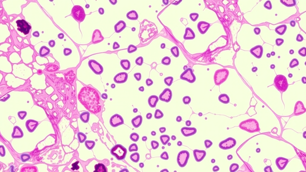
Breast cancer remains the most commonly diagnosed cancer in women worldwide and a leading cause of cancer-related death, and for more than a century the decisive diagnostic tool has been the trained human eye. Pathologists examine thin slices of tumor tissue stained with Hematoxylin and Eosin, searching for the subtle architectural and cellular fingerprints that separate a harmless lesion from a life-threatening carcinoma. It is a process that works, but it is slow, demanding, and notoriously vulnerable to variability between observers. Two experts looking at the same slide do not always agree, and with whole-slide digital scanners now flooding laboratories with gigantic images, the workload has become a genuine bottleneck in clinical care.
A team of researchers at the University of Naples Federico II—Antonio M. Rinaldi, Cristiano Russo and Cristian Tommasino—has now published a study in the journal Data Mining and Knowledge Discovery that tackles this problem not by making image-recognition models bigger, but by teaching them to see tissue the way biologists do: as networks of interacting cells and regions rather than as flat grids of pixels. Their framework, built on hierarchical graph neural networks, represents every detected cell nucleus as a node in a graph, every coherent tissue region as a node in a second, coarser graph, and then fuses the two levels into a single multi-scale structure that a neural network can reason over.
The motivation comes from a well-known weakness of the deep learning systems currently dominating computational pathology. Convolutional neural networks and Vision Transformers slice a digitized slide into fixed-size patches, extract local features from each patch, and aggregate the results into a final decision. This patch-based strategy delivers impressive accuracy, but it forces an awkward trade-off: high-resolution patches capture fine morphological detail but lose the surrounding context, while larger patches provide context at the cost of detail. More fundamentally, none of these architectures explicitly encodes the relationships between neighboring cells, the adjacency of tissue compartments, or the natural hierarchy that runs from nucleus to gland to tissue region—precisely the structures pathologists rely on when they reason through a diagnosis.
Graphs offer an escape from that limitation. In the Naples team’s pipeline, a Vision Transformer-based segmentation model first identifies individual nuclei within each tissue region of interest and produces pixel-wise embedding maps that encode local morphological and contextual information. Each nucleus becomes a node, described by features that can be handcrafted shape and texture descriptors, deep semantic vectors drawn from the segmentation network’s decoder, or a hybrid of both. Edges connect cells whose centroids lie close together, using a distance-constrained k-nearest-neighbor strategy in which the number of neighbors and a maximum distance threshold prevent biologically implausible long-range links. In parallel, a superpixel-based algorithm partitions the image into coherent tissue regions, and a Region Adjacency Graph links neighboring regions so that information can flow across shared biological interfaces.
The genuinely novel part is the hierarchical integration. A binary association matrix records which cell nodes belong to which tissue nodes, allowing structured information to propagate across scales. The researchers designed and compared three fusion architectures: a late-fusion model that processes cell and tissue graphs separately and concatenates the embeddings at the end; a layer-wise model that interleaves cellular and tissue information at every depth of the network; and a sequential model in which cellular information is fully processed first and then used to enrich tissue-level analysis, deliberately mimicking how a pathologist moves from the microscope to the wider field of view.
Which of these strategies wins was not obvious in advance, and the team treated the answer as an empirical question. They carried out a validation-driven Bayesian hyperparameter search across graph construction parameters, node feature configurations, pooling strategies, network depths, hidden dimensionality, and learning rates, always confining optimization to the validation split to avoid contaminating the test set. The systematic search covered k-nearest-neighbor values from 5 to 20, distance thresholds between 80 and 200 pixels, and graph network depths of two to four layers. Every reported result was averaged over multiple runs with different random seeds, with early stopping on validation loss to curb overfitting.
The evaluation was performed on BRACS, a clinically validated benchmark of breast carcinoma subtyping that stands out for its difficulty and realism. The dataset was collected at IRCCS Fondazione Pascale in Naples between 2019 and 2020, drawn from 189 patients aged 16 to 86, digitized at 40x magnification, and annotated by three board-certified pathologists using a collaborative review process to resolve disagreements. It contains 547 whole-slide images and 4,539 annotated regions spanning seven histological classes that trace the full progression of breast disease: normal tissue, pathological benign lesions, usual ductal hyperplasia, flat epithelial atypia, atypical ductal hyperplasia, ductal carcinoma in situ, and invasive carcinoma. Crucially, the data are split at the patient level, so no patient appears in more than one split—a design that prevents data leakage and reflects the reality of clinical deployment, where a model must generalize to people it has never seen.
The results, reported as weighted F1-scores to account for the dataset’s class imbalance, delivered a clear verdict: the sequential hierarchical fusion strategy proved most effective, achieving a weighted F1-score of 0.587 plus or minus 0.015 on the test set and outperforming both a reimplementation of the earlier HACT-NET baseline and the alternative fusion variants, while also showing lower variance. Along the way, the design-space analysis produced a set of practical lessons. Decoder-derived features from the segmentation network beat handcrafted descriptors; mean bounding-box pooling was more robust than contour-based pooling; the CellViT segmentation backbone yielded the most informative cellular features; and the best overall recipe paired Principal Neighborhood Aggregation layers for the cell graph with Graph Isomorphism Network layers for the tissue graph, which consistently outperformed PNA layers at the tissue level in both accuracy and stability.
Perhaps the most clinically significant finding is where the improvements concentrated. The hardest categories in breast histopathology are not the obvious ones—distinguishing invasive carcinoma from normal tissue is relatively straightforward—but the atypical and precancerous lesions that sit on the borderline between benign and malignant. Here the hierarchical framework shone, with per-class F1 gains of up to 0.187 for atypical ductal hyperplasia, the borderline lesion most associated with elevated carcinoma risk, along with consistent improvements for flat epithelial atypia and ductal carcinoma in situ. The confusion matrix showed that most remaining errors occurred between normal and pathological benign tissues, two non-malignant categories where a cautious misclassification carries far less clinical consequence than confusing an atypical lesion with a harmless one.
The authors are careful about the limits of their claims. The study is confined to supervised classification at the region-of-interest level on a single dataset; comparisons with large pathology foundation models and weakly supervised whole-slide pipelines are left to future work, as those address different problem formulations. The tissue regionalization relies on unsupervised superpixels, and segmentation and graph construction carry a nontrivial computational cost, although spatial indexing keeps neighborhood construction close to linear in practice for typical region sizes containing hundreds to a few thousand nuclei. The team also notes that while qualitative inspection of the learned embeddings showed atypical nuclei clustering with tissue regions exhibiting disrupted glandular organization—a pattern consistent with diagnostic criteria—expert-led validation of this interpretability remains follow-up work. Still, the framework’s modular design means it can be extended to whole-slide graphs, to other cancer types, and to emerging relational domains such as histogenomics and spatial transcriptomics. In an era when artificial intelligence is being asked to shoulder an ever-greater share of diagnostic labor, the Naples study makes a compelling argument that the path to trustworthy machine diagnosis runs not through bigger pixel-grinders, but through models that respect the biology written into the tissue itself.
Subject of Research: Hierarchical graph neural networks for classifying breast cancer subtypes from histopathological images
Article Title: Hierarchical graph networks for breast cancer subtype classification
Article References: Rinaldi, A. M., Russo, C., & Tommasino, C. (2026). Hierarchical graph networks for breast cancer subtype classification. Data Mining and Knowledge Discovery, 40(6), Article 104. https://doi.org/10.1007/s10618-026-01259-y
Image Credits: AI Generated
DOI: 10.1007/s10618-026-01259-y
Keywords: graph neural networks, breast cancer, digital pathology, histopathology, BRACS dataset, cell graphs, tissue graphs, deep learning, computer-aided diagnosis, hierarchical fusion, cancer subtyping, Vision Transformers
Cite Scienmag News
Nathaniel Bowman. (September 22, 2026). Graph neural networks teach AI to read breast cancer slides like pathologists. Scienmag. https://scienmag.com/graph-neural-networks-teach-ai-to-read-breast-cancer-slides-like-pathologists/
Nathaniel Bowman. "Graph neural networks teach AI to read breast cancer slides like pathologists." Scienmag, 22 September 2026, https://scienmag.com/graph-neural-networks-teach-ai-to-read-breast-cancer-slides-like-pathologists/. Accessed 5 October 2026.
Nathaniel Bowman. "Graph neural networks teach AI to read breast cancer slides like pathologists." Scienmag. September 22, 2026. https://scienmag.com/graph-neural-networks-teach-ai-to-read-breast-cancer-slides-like-pathologists/


