


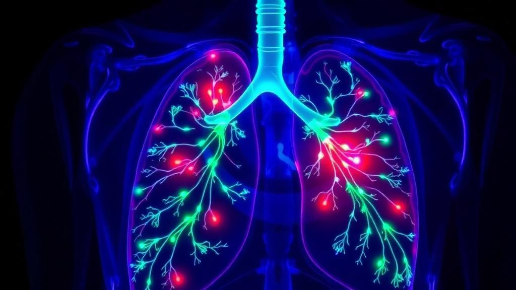
Lung cancer screening programs built around low-dose computed tomography are catching tumors earlier than ever before, and that success has created an unexpected technical problem. The nodules now being flagged on CT scans are often smaller than a centimeter, roughly the size of a pea, and once a patient is on the operating table, a surgeon peering through a video-assisted thoracoscope may simply not be able to see or feel them. A new study from Charité-Universitätsmedizin Berlin, published in CVIR Oncology, describes a practical solution: interventional radiologists mark the nodule with dye before surgery, using either a vivid blue stain or a fluorescent green beacon that shines through lung tissue under near-infrared light.
The report, led by interventional radiologist Charlie A. Hamm and colleagues, is framed as a technical roadmap rather than a large trial, but its results are striking in their consistency. Eleven patients, four women and seven men ranging in age from 34 to 77, underwent CT-guided dye-based marking of twelve lung nodules before minimally invasive, lung-sparing resection. The median nodule diameter was just 8.5 millimeters. Every single marking procedure was technically successful, every lesion was completely resected, and no operation had to be converted to open surgery or an entire lobectomy because the surgeon could not find the target. Seven of the twelve lesions turned out to be malignant, underscoring how much rides on getting the localization right.
The clinical backdrop explains why this matters. Randomized screening trials such as the Dutch-Belgian NELSON study showed that volume-based CT screening reduces lung cancer mortality, and more recent cohort work like the SUMMIT study has confirmed that the majority of screen-detected cancers are diagnosed at stage I or II, when surgery is the primary treatment. That shift has pushed thoracic surgeons toward sublobar resection, removing a wedge of lung or a single segment rather than the whole lobe, to preserve as much healthy parenchyma as possible. But the smaller the lesion and the more tissue-sparing the operation, the more critical it becomes to know exactly where the target sits before the first incision is made.
The traditional answer has been the hook-wire, a thin barbed wire placed through the chest wall into or next to the nodule on the morning of surgery. Hook-wires work, but they come with real drawbacks. They typically force same-day scheduling of two procedures, they can dislodge or migrate before the surgeon reaches them, and a rigid wire protruding through the pleura is uncomfortable and potentially hazardous for the patient, who must be transported carefully to avoid tearing lung tissue. The Berlin team’s dye-based approach eliminates the transpleural hardware entirely, which the authors note has been reported to cause less patient discomfort and stress and reduces the risk of marker displacement.
The first of the two dye techniques relies on patent blue V, a synthetic triphenylmethane dye that has long been used to trace lymphatic drainage in breast surgery and melanoma staging. It is water-soluble, renally excreted, and has a low toxicity profile, with allergic reactions occurring in fewer than one percent of patients. Under CT-fluoroscopic guidance, the radiologist advances a fine 22-gauge Chiba needle until its tip sits immediately adjacent to the nodule, deliberately avoiding puncture of the lesion itself, which reduces the theoretical risk of tumor seeding along the needle track and avoids compromising the pathology specimen. Then 0.1 to 0.4 milliliters of dye is injected through a small Luer-lock syringe, and the needle is withdrawn promptly without a saline flush, which would dilute the dye and could tattoo the skin along the exit path.
What happens next is a matter of physics and anatomy. The dye diffuses through the terminal vascular bed of the lung and reaches the pleural surface, where it creates a sharply demarcated blue patch that tells the surgeon precisely where the resection margin should fall. If the lesion lies more than three centimeters beneath the pleura, the dye is injected in increments as the needle is withdrawn, leaving a stained tract running from the nodule toward the chest wall. Because patent blue V gradually disperses into the bloodstream and pleural fluid, the color fades within hours, so surgery must be scheduled the same day, ideally within three hours of the marking. In the Berlin series, the five patients who received patent blue V injections had a median marking time of just seven minutes.
The second technique solves the fading problem with indocyanine green, or ICG, a tricarbocyanine fluorescent dye that binds readily to tissue proteins and glows bright emerald green when excited by near-infrared light. Injected alone, ICG diffuses more slowly than patent blue V and can remain visible in lung tissue for up to six days, a finding documented in prior experimental work. More importantly, near-infrared light penetrates soft tissue far better than visible light, so a fluorescence camera in the operating room can detect the glowing signal from millimeters to several centimeters beneath the pleural surface, effectively letting the surgical team see through the lung’s outer rim to a depth of roughly three centimeters. The six patients in the series who received ICG-based marking had a median procedure time of 16.5 minutes.
The most innovative variant described in the report goes one step further: instead of injecting liquid dye that will eventually disperse, the team deploys a tiny embolization coil soaked in ICG. The preparation is meticulous. ICG is mixed with ten milliliters of sterile water, and two to three milliliters of the solution are used to slowly flush the coil introducer outside the patient to prevent premature deployment. The markers are Tornado-type coils measuring 0.035 inches in diameter and 7 to 8 millimeters in length. After percutaneous puncture with an 18-gauge coaxial needle, the coil is advanced with the stiff end of a guidewire and deployed under CT-fluoroscopy immediately adjacent to the lesion, with care taken not to push it too far past the target. The coil acts as a physical scaffold that holds the dye in place, and because the fluorescence persists for days, surgery can be scheduled up to thirteen days after marking, giving hospitals valuable flexibility when operating rooms are crowded or schedules shift unexpectedly.
Safety data from the series align with the broader literature. Only one complication occurred, an asymptomatic pneumothorax during needle puncture that was managed conservatively without any intervention, consistent with reported pneumothorax rates of around six percent for CT-guided ICG injection in a retrospective analysis of 471 patients. The authors do caution that coil placement requires a larger 18-gauge needle than pure dye injection, which may influence complication rates. Prior studies have reported technical success rates above 98 percent for CT-guided dye injection and 100 percent for bronchoscopy-guided ICG-coil placement, and the addition of preoperative markings has been shown in other reports to drive conversion rates to open surgery close to zero.
What makes the Berlin report notable is the delivery route. ICG-soaked coil marking had previously been described through bronchoscopy, a technique that requires general anesthesia and specialized endoscopic equipment, and only recently through a percutaneous approach. The CT-guided percutaneous route can be performed in an outpatient workflow under local anesthesia alone, making it less resource-intensive and easier to slot into a busy hospital schedule. The authors emphasize that the technique is ultimately a story about multidisciplinary collaboration: the tumor board decides the patient needs lung-sparing surgery, the interventional radiologist plants the beacon, and the thoracic surgeon follows the glow. As robotic-assisted and uniportal thoracoscopic surgery become more common and screening programs continue to surface ever-smaller nodules, dye-based marking may become a routine bridge between the radiology suite and the operating room, turning invisible targets into ones a surgeon cannot miss.
Subject of Research: CT-guided preoperative dye-based marking of small lung nodules for minimally invasive surgical resection
Article Title: CT-guided percutaneous marking of lung nodules using patent blue V and indocyanine green
Article References: Hamm, C. A., Elsner, A., Fehrenbach, U., Savic, L. J., Bawaadam, H., Collettini, F., Neudecker, J., Gebauer, B., Rückert, J., & Auer, T. A. (2026). CT-guided percutaneous marking of lung nodules using patent blue V and indocyanine green. CVIR Oncology, 2(1), Article 2. https://doi.org/10.1007/s44343-026-00031-9
Image Credits: AI Generated
DOI: 10.1007/s44343-026-00031-9
Keywords: lung nodules, interventional radiology, indocyanine green, patent blue V, CT guidance, thoracic surgery, fluorescence imaging, preoperative localization, lung cancer screening, video-assisted thoracoscopic surgery, fiducial coils, lung-sparing resection
Cite Scienmag News
Nathaniel Bowman. (September 30, 2026). Glowing Dyes and Tiny Coils Help Surgeons Find Hidden Lung Nodules. Scienmag. https://scienmag.com/glowing-dyes-and-tiny-coils-help-surgeons-find-hidden-lung-nodules/
Nathaniel Bowman. "Glowing Dyes and Tiny Coils Help Surgeons Find Hidden Lung Nodules." Scienmag, 30 September 2026, https://scienmag.com/glowing-dyes-and-tiny-coils-help-surgeons-find-hidden-lung-nodules/. Accessed 5 October 2026.
Nathaniel Bowman. "Glowing Dyes and Tiny Coils Help Surgeons Find Hidden Lung Nodules." Scienmag. September 30, 2026. https://scienmag.com/glowing-dyes-and-tiny-coils-help-surgeons-find-hidden-lung-nodules/


