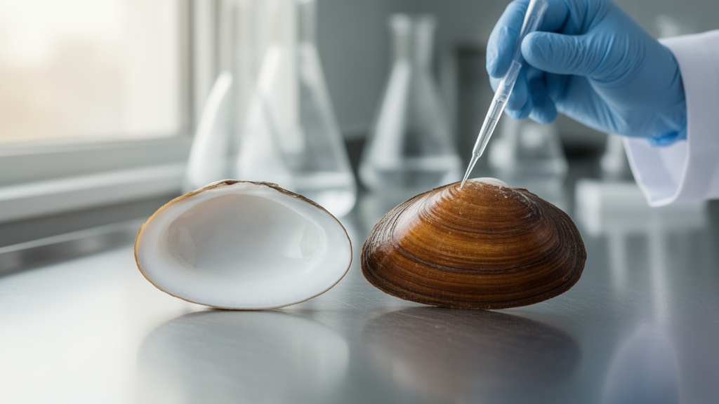
Scientists have shown that silencing a single gene in the dwarf surf clam can transform its uniformly white shell into a brownish-yellow one, simply by allowing a natural pigment to build up inside the animal. The gene, called FECH, encodes ferrochelatase, the enzyme that performs the final step of heme synthesis by inserting iron into protoporphyrin IX, or PPIX. When that step is blocked, PPIX accumulates, and because PPIX is naturally orange-brown, the color becomes visible in the shell and internal organs without any dyes, substrates, or equipment. The study, published in BMC Marine Science, was led by Dongxue Su, Ang Zhao, and colleagues, and describes a proof-of-concept route toward using shell color as a visible reporter in bivalves.
The motivation comes from a practical gap in molluscan genetics. Reporter systems such as green fluorescent protein require excitation light and are vulnerable to tissue autofluorescence, while enzyme reporters like β-glucuronidase need costly substrates and usually demand invasive sampling or tissue dissection. In mollusks, where gene editing tools are still maturing, visible phenotypic markers for confirming transgene integration are scarce. Shell color offers an attractive alternative because it is observable from the outside, continuously, and at essentially no cost. The problem for the dwarf surf clam specifically is that, unlike many bivalves, it naturally produces extremely uniform white shells, so its color cannot serve as a marker without first inducing pigment accumulation.
The team began with feeding experiments. Thirty-day-post-fertilization clams were divided into groups receiving either plain microalgae or microalgae mixed with 0.01 percent by weight of one of three pigments commonly used as aquaculture feed additives: astaxanthin, hemin, or chlorophyllin. Each treatment was run in three independent aquaria of 45 individuals, with the pigment mixture fed for 20 days followed by a 10-day depuration period on pure microalgae to flush out unabsorbed residue. After the feeding phase, the astaxanthin group showed an orange-yellow shell, the hemin group an orange-red shell, and the chlorophyllin group a bluish-green shell.
Dissection revealed visible pigment accumulation in the visceral mass, which appeared darker than the shell, with hemin producing the most pronounced effect. Enzyme-linked immunosorbent assay, or ELISA, measurements of pigment content in the gill, mantle, digestive gland, muscle, and foot tissues showed that all three treated groups had higher pigment levels than controls, particularly in the gill, mantle, and digestive gland, with hemin producing a significantly greater increase than the other pigments. The statistical unit was the aquarium, not the individual clam: two individuals per aquarium were sampled and averaged, giving three independent replicates per treatment per tissue per time point. The authors note that hemin is the stable oxidized, chloride-coordinated form of heme, which is itself a product of porphyrin metabolism, so the strongest response to hemin pointed them directly toward the porphyrin and heme biosynthesis pathway.
Searching the M. lateralis genome, the researchers identified five candidate genes in that pathway: PBGD, UROS, UROD, TSPO, and FECH. Sequence analysis confirmed that the clam FECH gene contains nine exons and eight introns and carries the conserved Ferrochelatase and ZnF_C2H2 domains, with all known functional amino acid residues intact, including the catalytic histidine and glutamate pair, the methionine involved in iron binding, and the metal-sensitive QSK loop that regulates product release. Phylogenetic analysis placed the bivalve FECH sequences in a single well-supported lineage consistent with known species relationships, supporting evolutionary conservation of the enzyme’s function across animals.
Transcriptome data across eleven developmental stages and seven adult tissues showed that FECH is expressed consistently from the trochophore stage through the spat stage and maintains high expression across adult tissues, with the highest overall expression level among the five candidate genes in adult tissues. Because FECH’s substrate is PPIX, which imparts a reddish-brown color when it is not metabolized to heme, the authors interpreted this sustained expression as suggesting a regulatory role in PPIX homeostasis and shell pigment deposition. They caution that this is an association drawn from expression patterns; the functional test came next.
For the functional experiment, the team used RNA interference. They designed a target sequence with bioinformatic screening against the clam genome and transcriptome to minimize off-target effects, cloned it into the L4440 vector, and produced double-stranded RNA in HT115 bacteria, which were fed to the clams mixed with microalgae. Thirty-day-post-fertilization clams were divided into three groups: a plain microalgae control, an empty-vector control, and the FECH-RNAi group, each with three aquarium-level replicates of 45 individuals. After one month of treatment, FECH expression was significantly downregulated in the gill, mantle, and digestive gland tissues, and the shell color of the RNAi group had shifted from white to brownish-yellow, with a measured RGB value of 113, 94, 71. The visceral mass turned reddish-brown, with an RGB value of 116, 71, 59.
The molecular measurements matched the visible phenotype. ELISA assays showed that heme content decreased significantly in the gill, mantle, and digestive gland of the RNAi group, while ELISA-detected PPIX accumulated significantly in those same tissues, both at the p < 0.05 threshold using the Kruskal-Wallis test followed by Dunn’s post-hoc test. The authors are careful in their language: they describe these results as correlative evidence that FECH participates in modulating PPIX accumulation dynamics and as regulatory involvement in the color phenotypes, rather than a fully established causal chain. They point out that in the Pacific oyster, a previous study found that knockdown of the FECH ortholog increases PPIX content and deepens orange shell coloration, a result consistent with what they observed in the clam.
The proposed application is an endogenous, non-invasive visible reporter system. Because PPIX is naturally orange-brown and can be seen without any equipment or exogenous substrates, the FECH/PPIX axis could in principle allow researchers to track gene expression or visualize gene editing events in M. lateralis, a species valued as the molluscan equivalent of the fruit fly for its fast growth, roughly two-month generation time, and high fecundity. The authors stress, however, that this remains a proof of concept. The RNAi experiment covered a single developmental stage and one month of observation, so long-term stability of FECH inhibition and applicability across the full life cycle remain untested. Direct genetic evidence, such as CRISPR-Cas9 knockout of FECH, would strengthen the causal interpretation, and whether the system works in other bivalve species is unknown.
There are also open biological questions. Other porphyrin pathway genes may contribute to shell coloration: UROS, a rate-limiting enzyme in early porphyrin synthesis, is highly expressed during early developmental stages, and TSPO, a mitochondrial porphyrin transporter, is expressed from the trochophore stage onward and might influence PPIX localization within cells. The authors suggest that future work should construct stable transgenic clam lines with tissue-specific or inducible FECH expression, dissect the independent and combined roles of the other pathway genes through multi-gene interference, and carry out long-term phenotyping and cross-species validation before any practical use. Within the scope of the genes examined, the study establishes that reducing FECH activity in the dwarf surf clam lowers heme, raises detectable PPIX, and visibly recolors the shell, providing a foundation for a simple and economical reporter that could eventually help researchers isolate gene-edited bivalves by eye.
Subject of Research: FECH regulation of protoporphyrin IX accumulation and shell coloration in the dwarf surf clam Mulinia lateralis
Article Title: FECH negatively regulates protoporphyrin IX accumulation and shell coloration in the dwarf surf clam Mulinia lateralis
Article References: Su, D., Zhao, A., Wang, H., Li, C., Bao, L., Zhu, T., Huang, X., Yang, Z., & Bao, Z. (2026). FECH negatively regulates protoporphyrin IX accumulation and shell coloration in the dwarf surf clam Mulinia lateralis. BMC Marine Science, 1(1), Article 9. https://doi.org/10.1186/s44479-026-00011-2
Image Credits: AI Generated
DOI: 10.1186/s44479-026-00011-2
Keywords: FECH, protoporphyrin IX, shell color, Mulinia lateralis, bivalve, RNA interference, heme biosynthesis, porphyrin pathway, reporter system, pigment accumulation, ELISA, marine genetics
Cite Scienmag News
Juliet Wilcox. (October 27, 2026). Gene knockdown turns white clam shells brown in new study. Scienmag. https://scienmag.com/gene-knockdown-turns-white-clam-shells-brown-in-new-study/
Juliet Wilcox. "Gene knockdown turns white clam shells brown in new study." Scienmag, 27 October 2026, https://scienmag.com/gene-knockdown-turns-white-clam-shells-brown-in-new-study/. Accessed 28 October 2026.
Juliet Wilcox. "Gene knockdown turns white clam shells brown in new study." Scienmag. October 27, 2026. https://scienmag.com/gene-knockdown-turns-white-clam-shells-brown-in-new-study/