

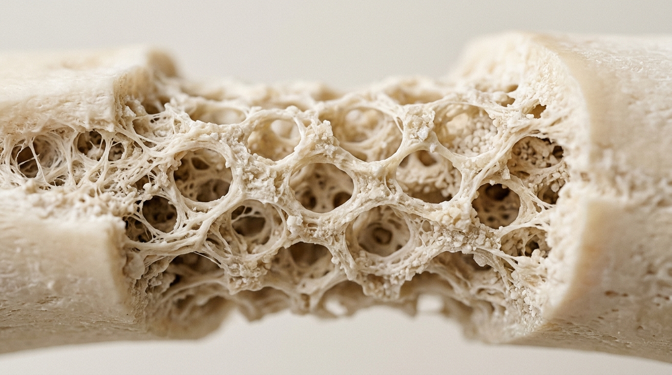
Gene-activated matrices represent a converging field of nanomedicine and biomaterials engineering designed to overcome the biological limitations of critical-size bone defects. A comprehensive narrative review published in Materials Today Bio examines how these platforms integrate structural scaffolds with nucleic acid delivery to sustain osteogenic signaling throughout the repair process. The study identifies a fundamental mismatch in current orthopedic treatments: while bone possesses the capacity for scarless regeneration, defects exceeding a specific size threshold cannot be bridged by endogenous signaling alone. This review traces the technical evolution of these systems from scaffold bioactivity to clinical translation, highlighting the specific pharmacokinetic advantages of gene-based strategies over traditional protein biologics.
The primary challenge in bone regeneration is the duration of therapeutic signaling rather than its magnitude. Critical-size defects, commonly defined as a loss of at least half the bone circumference over a length greater than 1 to 2 cm, arrest spontaneous repair. Traditional clinical benchmarks, such as autografts, are limited by finite harvest volume and donor site morbidity. Allografts and xenografts expand supply but lose viable osteogenic cells during processing, leaving them as passive conduits rather than active instructional agents. Protein biologics, while potent, are transient; their short half-life at the defect site necessitates supraphysiological dosing, which increases the risk of ectopic bone formation and inflammatory reactions.
Gene-activated matrices address this limitation by delivering the genetic instructions for therapeutic proteins rather than the proteins themselves. By embedding nucleic acid cargo within a biomaterial scaffold, these platforms turn resident host cells into local bioreactors. This approach ensures that osteogenic signals are produced endogenously at physiologically regulated concentrations for a duration governed by the scaffold’s degradation kinetics. The review emphasizes that the scaffold is not an inert carrier but an active component of the delivery system, where surface chemistry, porosity, and mechanical properties directly influence vector retention and cellular transfection efficiency.
The review details the specific material classes that serve as the foundation for these matrices. Bioceramics, such as calcium phosphate and bioactive glass, act through ionic dissolution and mineral affinity, which can both enhance osteoinduction and sequester nucleic acid vectors. Natural polymers like collagen and chitosan provide cell-recognition motifs that facilitate integrin-mediated signaling, while synthetic polymers such as PLGA and PCL offer tunable degradation rates. The choice of material is critical because it determines the local microenvironment, including the balance between pro-inflammatory M1 and pro-regenerative M2 macrophage phenotypes, which is essential for successful tissue integration.
Delivery systems for nucleic acids vary significantly in their transduction efficiency, expression duration, and immunogenicity. Viral vectors, including adenoviral and lentiviral systems, offer high-efficiency delivery but face constraints from pre-existing immunity and the risk of insertional mutagenesis. Non-viral platforms, such as lipid nanoparticles and polymeric polyplexes, avoid integration risks but often suffer from lower transfection rates and rapid clearance from the bone-poor vasculature. Calcium phosphate nanoparticles provide a unique advantage by aligning carrier function with therapeutic osteogenicity, though they are limited by aggregation and variable nucleic acid loading. The review notes that no single vector class is optimal across all bone repair applications, necessitating a tailored approach based on the specific clinical context.
The design of gene-activated matrices involves complex loading and release architectures that must be synchronized with the biological phases of repair. Surface adsorption offers simplicity but risks burst release, while bulk encapsulation and nanoparticle immobilization provide more sustained delivery profiles. The 3D microenvironment of the scaffold plays a decisive role in transfection, as matrix stiffness and surface biochemistry can enhance or inhibit the internalization of gene delivery vectors. For instance, studies have shown that 3D confinement in collagen-alginate hydrogels can significantly amplify osteogenic marker expression compared to monolayer cultures, demonstrating that the physical context of the scaffold is as important as the genetic cargo itself.
Multi-factor delivery systems are emerging as a key frontier in GAM engineering. Rather than delivering a single osteogenic signal, these platforms co-deliver multiple nucleic acid cargoes to coordinate angiogenesis and osteogenesis. Sequential delivery of vascular endothelial growth factor followed by bone morphogenetic protein-2 has been shown to outperform simultaneous delivery in preclinical models. This temporal coordination mimics the natural progression of bone repair, where vascular invasion must precede and support mineralization. The review highlights that the spatial and temporal arrangement of these signals within the scaffold is a primary determinant of the final regenerative outcome.
The intellectual property landscape for gene-activated matrices reveals a significant gap between scientific output and patent protection. Most foundational constructs remain unpatented or are covered only by academic institutional filings, while large orthobiologics manufacturers hold dominant positions in protein-based delivery. A few key patents define the current commercial space, including a Russian SME patent for patient-specific 3D-printed calcium phosphate scaffolds and several US grants for lentiviral cell-based regional gene therapy. However, substantial white space remains, particularly for non-viral mRNA delivery within 3D scaffolds and stimulus-responsive release systems, which are currently unprotected and represent significant opportunities for future innovation.
Despite preclinical promise, clinical translation faces substantial translational barriers. Manufacturing scalability is a major challenge, as cell-based GAMs require complex ex vivo processing that is costly and variable. Sterilization methods must be carefully selected to avoid damaging the nucleic acid cargo or the scaffold architecture. Regulatory classification is also complex, as these products are often treated as combination products where the gene therapy component drives the primary mode of action. To date, only a small number of human studies have been conducted, including a maxillofacial trial that reported no adverse events over 30 months and a spinal fusion study that showed comparable outcomes to autograft, though the latter was not designed to demonstrate superiority.
The review concludes that the gap between preclinical validation and clinical deployment is not narrowing at a pace that reflects mechanistic progress. Most evidence derives from rodent models, which cannot fully reproduce the geometric and immunological complexity of human skeletal repair. Future directions include the integration of CRISPR-based gene regulation, stimuli-responsive fabrication, and computational design to optimize the spatiotemporal control of gene expression. While gene-activated matrices have advanced from proof-of-concept to sophisticated nanomedicine platforms, they remain largely in the preclinical or early investigational phase, requiring further advances in manufacturing and large-animal validation before routine clinical use can be achieved.
The review highlights that scaffold surface chemistry directly influences the immune response at the defect site, a factor critical for successful tissue integration. Physicochemical properties such as surface topography, wettability, and stiffness regulate macrophage polarization, determining whether healing progresses toward tissue formation or fibrous encapsulation. Successful repair requires a controlled transition from the pro-inflammatory M1 phenotype, which drives early inflammation through cytokines like TNF-α and IL-1β, to the pro-regenerative M2 phenotype, which resolves inflammation via IL-10. Scaffold design can actively engineer this transition; for instance, nano-scale hydroxyapatite particles have been shown to induce M2 polarization through the STAT3/c-Maf axis, thereby promoting osteogenesis and endothelial tube formation. This immunomodulatory capacity is a first-class design parameter for gene-activated matrices, as cell-based constructs have demonstrated the ability to generate M2-skewed macrophage populations more effectively than recombinant protein controls.
Specific osteogenic gene targets have shown varying degrees of efficacy in preclinical models, with bone morphogenetic protein-2 (BMP-2) producing the most consistent evidence of in vivo bone formation. The shift from plasmid DNA to chemically modified mRNA (cmRNA) has sharpened delivery efficacy and safety. In critical-sized femoral defects, BMP-2 cmRNA achieved complete defect bridging at a 50 μg dose, avoiding the excessive bone formation and delayed mechanical recovery associated with recombinant BMP-2. This nucleic acid-mediated approach matches recombinant protein outcomes at substantially lower doses, though optimal carrier formulation and defect geometry remain incompletely characterized. Vascular endothelial growth factor (VEGF) functions as a mechanistically coupled co-target, where its osteogenic contribution depends on coordinated BMP-2 co-expression rather than expression magnitude alone. Dual-gene scaffolds have demonstrated that high transgene output does not always correlate with higher mineralization, indicating that the spatial and temporal coordination of signals is more critical than the sheer volume of protein produced.
Preclinical model limitations significantly constrain the interpretation of these findings. Small rodent models, such as rat calvarial defects, heal through intramembranous ossification across a thin cortical layer flanked by vascular tables, a process that lacks the axial load, functional periosteum, and marrow cavity vascular invasion required for long bone segmental repair. Critical size is a property of the specific model rather than a fixed dimension, with reported rat calvarial thresholds generally falling between 5 and 8 mm. For load-bearing evaluation, sheep femoral mid-diaphyseal osteotomies have been proposed as a standardizable large-animal system, yet no gene-activated matrix construct has been tested in this model at clinically relevant scale. The preferential publication of positive findings and inadequate reporting of randomization and blinding in some preclinical studies may contribute to an overestimation of efficacy, limiting the reliability of cross-study comparisons.
Manufacturing and sterilization present distinct challenges depending on whether the construct is acellular or cell-based. Acellular constructs can be produced in advance and implanted in a single operation, relying on host cells migrating into the implant. In contrast, cell-based designs require ex vivo transduction and expansion, introducing donor variability and high manufacturing costs estimated at $100,000 to $300,000 per patient. Sterility assurance has no generic solution; terminal methods like autoclaving or gamma irradiation can alter particle size or damage cargo, while sterile filtration faces yield constraints due to the small diameter of mRNA-lipid nanoparticles. Storage stability is equally formulation-specific, with conventional cryoprotectants retaining only 60–70% of transfection activity after four weeks at −80 °C in one mRNA-LNP model. These technical adaptations are sequential, meaning a formulation that loses activity during sterilization never reaches stability studies, and a trial lacking a scaffold-only arm cannot attribute its outcome to the transgene.
Subject of Research: Gene-activated matrices as nanomedicine platforms for bone regeneration
Article Title: Gene-activated matrices as nanomedicine platforms for bone regeneration: Delivery, translation and patent landscape
Article References: Abdelnabi, H., Alkaabi, S. M., Alderei, K. S., Alaryani, A. S., & Mohsin, S. (2026). Gene-activated matrices as nanomedicine platforms for bone regeneration: Delivery, translation and patent landscape. Materials Today Bio, 41, Article 103808. https://doi.org/10.1016/j.mtbio.2026.103808
Image Credits: AI Generated
DOI: 10.1016/j.mtbio.2026.103808
Keywords: bone regeneration, nanomedicine, gene therapy, biomaterials, tissue engineering, patent landscape, scaffold design, osteogenesis, Gene-activated, matrices, platforms, bone
Cite Scienmag News
Gregory Coleman. (November 8, 2026). Gene-Activated Matrices Bridge Bone Repair Gap. Scienmag. https://scienmag.com/gene-activated-matrices-bridge-bone-repair-gap/
Gregory Coleman. "Gene-Activated Matrices Bridge Bone Repair Gap." Scienmag, 8 November 2026, https://scienmag.com/gene-activated-matrices-bridge-bone-repair-gap/. Accessed 8 November 2026.
Gregory Coleman. "Gene-Activated Matrices Bridge Bone Repair Gap." Scienmag. November 8, 2026. https://scienmag.com/gene-activated-matrices-bridge-bone-repair-gap/