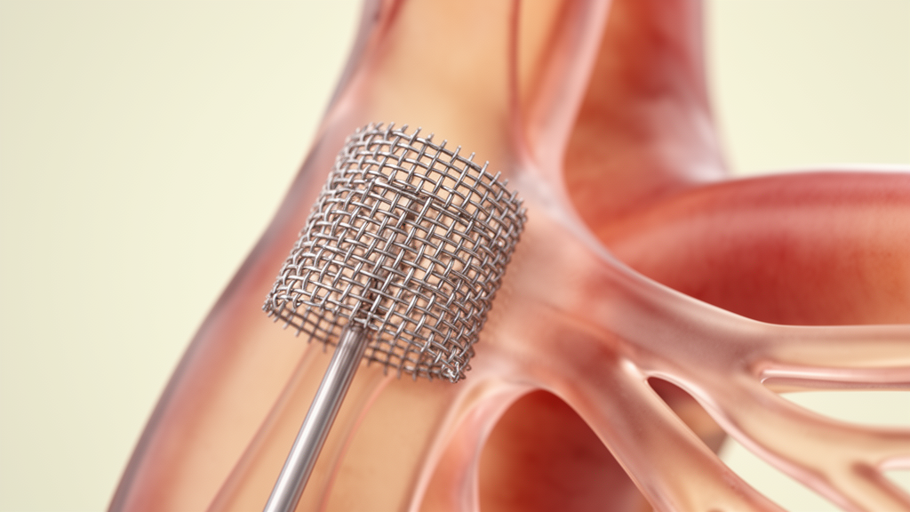
Endovascular specialists have spent the past fifteen years building a better toolkit for reopening chronically blocked subclavian arteries, but a new review reaches a sobering conclusion: the tools have multiplied while the proof of benefit has not. Writing in CVIR Endovascular, researchers led by Srđan Babić and Milovan Bojić trace how the treatment of chronic total occlusion of the subclavian artery has evolved since one of the earliest dedicated long-term series was published in 2012, and they find that technological progress has broadened treatment options without clearly improving technical success or long-term patency.
The subclavian artery supplies the arm and, through the vertebral artery, contributes blood flow to the back of the brain. When it becomes completely and chronically blocked, the consequences can be serious: vertebrobasilar insufficiency, ischemia of the upper extremity, coronary-subclavian steal syndrome, and compromised inflow through the left internal mammary artery in patients who have undergone coronary artery bypass grafting. Surgical reconstruction was historically considered the gold standard, but over the last two decades endovascular treatment has become the usual first consideration in appropriately selected symptomatic patients, even though comparative evidence specific to complete occlusions remains limited.
The 2012 benchmark series, published online in March 2011, reported successful recanalization in 46 of 56 patients, a technical success rate of 82.1 percent, with primary patency of 97.9 percent at one year and 82.7 percent at three years. What makes that report remarkable is that it was achieved without any of the devices now considered standard. Dedicated peripheral chronic total occlusion devices did not exist, so operators relied on conventional 0.014-inch coronary guidewires, coronary guiding catheters, balloon angioplasty, and first-generation balloon-expandable or self-expanding stents. Subintimal recanalization was deliberately avoided, with repeated attempts made to achieve true intraluminal passage.
That early series also anticipated its own obsolescence. The authors predicted that future improvements in technical success would depend on dedicated guidewires designed specifically for crossing chronic occlusions and on more accurate preprocedural imaging for lesion characterization. Fifteen years later, the review confirms, both predictions have largely come true. Contemporary practice now includes dedicated chronic total occlusion guidewires with varied tip loads, enhanced torque control, and hydrophilic or polymer-jacketed coatings; microcatheters and support catheters; refined bidirectional crossing strategies; low-profile delivery systems; advanced stent platforms; high-resolution computed tomography angiography; and selective adjunctive intravascular imaging.
Imaging has changed particularly markedly. In the original cohort, digital subtraction angiography was used in 15 patients between 1999 and 2005, after which multislice computed tomography angiography was introduced and applied in 41 patients. Today’s multidetector computed tomography angiography with multiplanar reconstruction permits detailed assessment of occlusion length, calcification, stump morphology, vessel tortuosity, the vertebral artery origin, and distal vessel quality before the first incision or puncture. Yet the review is explicit that whether routine advanced imaging actually improves technical success, safety, or patency in subclavian occlusion has never been established by comparative studies.
The published outcome data reinforce that caution. Across reported series, technical success has varied considerably: 82.1 percent in the original 2012 cohort, 77.6 percent in a study by Liu and colleagues, 93.8 percent in a series by Çakar and colleagues, and 91.3 percent in a series by Niu and colleagues. Differences in patient selection and lesion characteristics make direct comparison between these studies unreliable. Liu and colleagues also included a nonrandomized comparison with 21 patients treated by bypass surgery, in whom technical success was 100 percent and three-year primary patency was 92.1 percent after successful endovascular treatment versus 93.1 percent after surgery — results the review stresses cannot establish the superiority of either approach because treatment allocation was not randomized.
Newer adjunctive technologies show a similar pattern of promise without proof. Intravascular ultrasound may assist vessel sizing and evaluation of stent expansion, but the observational evidence includes predominantly stenotic lesions and does not establish a benefit specific to complete occlusions. Distal embolic protection has been described in a four-patient series of chronic brachiocephalic and subclavian occlusions with three successful recanalizations, demonstrating feasibility but not a reduction in cerebral embolic events. Calcium-modification techniques such as intravascular lithotripsy and orbital atherectomy have been reported in small series and isolated cases, including a case of severe radiation-associated subclavian disease with balloon underexpansion, but these reports do not establish improved crossing success, procedural safety, or long-term patency.
Complication data from the published series underscore that more specialized devices do not eliminate risk. The original 2012 cohort reported an overall complication rate of 7.1 percent. Sakai and colleagues reported four procedure-related events during 29 procedures, including two asymptomatic stent migrations, one hemianopsia, and one transient ischemic attack, along with three target-lesion reinterventions related to subacute thrombosis, in-stent restenosis, or stent-edge dissection. Niu and colleagues described a dissection extending toward the aortic arch that required covered-stent implantation, with subsequent compromise of flow at the vertebral artery origin. In contrast, Çakar and colleagues reported no periprocedural complications in 16 patients and one asymptomatic restenosis during two-year follow-up.
To illustrate the technological shift concretely, the review presents two case examples. In March 2008, a 22.5-millimeter occlusion of the proximal left subclavian artery was crossed with a 0.014-inch Miracle 3 guidewire supported by a 2.0 by 20-millimeter Maverick over-the-wire balloon catheter, followed by deployment of an 8.0 by 40-millimeter Wallstent. In April 2025, a short-segment occlusion of approximately 10 millimeters was crossed with a 0.018-inch ASAHI Halberd guidewire supported by a NaviCross catheter, predilated with a 5.5 by 20-millimeter Sterling balloon, and treated with a 7.0 by 39-millimeter Omnilink Elite balloon-expandable stent delivered through a Destination guiding sheath. The authors are careful to note that neither procedure, nor the comparison between two anatomically different cases, establishes improved technical success, safety, or patency.
What has actually changed, the review concludes, is the range of equipment rather than the fundamentals of the procedure. The essential decisions still concern lesion anatomy, safe wire passage, and appropriate stent placement, and the basic principles — adequate catheter support, controlled guidewire manipulation, confirmation of wire position, and appropriate stent selection — remain largely unchanged. Long, heavily calcified occlusions, flush ostial lesions, and those involving the vertebral artery origin remain technically difficult or unsafe, and their independent predictive value for technical failure has not been established in the available small series. For symptomatic patients in whom endovascular treatment is unsuitable or fails, carotid-subclavian bypass and subclavian-carotid transposition remain important alternatives. The authors call for prospective multicenter studies with standardized definitions of technical success, patency, and complications to determine whether further technological advances translate into meaningful clinical benefit for patients with subclavian artery chronic total occlusion.
Subject of Research: Technological evolution of endovascular recanalization of chronic total occlusion of the subclavian artery
Article Title: Endovascular recanalization of chronic total occlusion of the subclavian artery: fifteen years of technological evolution
Article References: Babić, S., Milačić, A., Kovačević, V., Pešić, S., Mihajlović, V., Antonić, Ž., & Bojić, M. (2026). Endovascular recanalization of chronic total occlusion of the subclavian artery: fifteen years of technological evolution. CVIR Endovascular, 9(1), Article 137. https://doi.org/10.1186/s42155-026-00793-6
Image Credits: AI Generated
DOI: 10.1186/s42155-026-00793-6
Keywords: subclavian artery, chronic total occlusion, endovascular treatment, guidewires, stents, computed tomography angiography, patency, technical success, intravascular lithotripsy, carotid-subclavian bypass, peripheral intervention, Endovascular
Cite Scienmag News
Ophelia Keating. (October 27, 2026). Fifteen Years of Subclavian Artery Occlusion Devices Have Not Yet Proven Better Outcomes. Scienmag. https://scienmag.com/fifteen-years-of-subclavian-artery-occlusion-devices-have-not-yet-proven-better-outcomes/
Ophelia Keating. "Fifteen Years of Subclavian Artery Occlusion Devices Have Not Yet Proven Better Outcomes." Scienmag, 27 October 2026, https://scienmag.com/fifteen-years-of-subclavian-artery-occlusion-devices-have-not-yet-proven-better-outcomes/. Accessed 28 October 2026.
Ophelia Keating. "Fifteen Years of Subclavian Artery Occlusion Devices Have Not Yet Proven Better Outcomes." Scienmag. October 27, 2026. https://scienmag.com/fifteen-years-of-subclavian-artery-occlusion-devices-have-not-yet-proven-better-outcomes/