


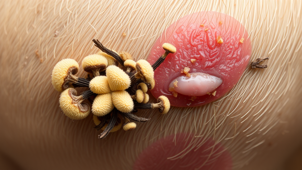
A fungal species better known for living quietly in soil and compost heaps has surfaced in an unexpected place: the inflamed skin of a family dog. In a case report published in Veterinary Medicine and Science, researchers at the University of Tehran describe what they believe is a rare recovery of Corynascus verrucosus, an environmental fungus that has never been firmly established as a pathogen in veterinary or human medicine, from a cutaneous lesion on a four-year-old spayed female Shih Tzu. The finding is less a cause for alarm than a demonstration of how modern molecular tools are reshaping the way veterinary diagnosticians identify the organisms behind unusual skin disease, and how cautious scientists must be when an environmental contaminant appears in a clinical sample.
The dog was referred to the Veterinary Clinic of the Faculty of Veterinary Medicine at the University of Tehran with a striking dermatological picture. It lived outdoors under unsanitary conditions with regular exposure to vegetation and soil, and it presented with diffuse hair loss, marked redness, itching and scaling across the skin of its back. Crucially, there was no ulceration or open wound, no lethargy and no loss of appetite, so systemic illness seemed unlikely and a complete blood count was not performed. The owner declined a skin biopsy, which meant the team could not obtain histopathological evidence of fungal invasion, a detail that would later shape how carefully the conclusions had to be framed. Hair plucks and skin scrapings were collected from the affected areas under sterile conditions and sent to the laboratory for fungal examination.
The initial diagnostic workup followed the standard playbook for suspected dermatophytosis, the superficial fungal infection of keratinized skin that is usually caused by organisms such as Microsporum canis, Microsporum gypseum or Trichophyton mentagrophytes. A Wood’s lamp examination was negative, and a direct microscopic examination of material treated with 10 percent potassium hydroxide also failed to reveal fungal elements. But the story changed when the samples were cultured on Sabouraud Dextrose Agar. After incubation at 28 degrees Celsius, colonies emerged that were creamy-brown and powdery with a colorless reverse surface. Lactophenol cotton blue staining of the growth revealed thin, septate hyphae studded with large, round, warty conidia, a morphological signature that immediately suggested the team was not dealing with a routine dermatophyte.
Seeking to probe the organism’s thermal behavior, the researchers subcultured the colonies onto blood agar and incubated them at 37 degrees Celsius, close to mammalian body temperature, for five days. The fungus produced creamy, dome-shaped, mucoid colonies under these conditions, and Giemsa staining revealed round-to-oval yeast-like cells with budding and point attachments when viewed under oil immersion at 100 times magnification. This dimorphic tendency, the ability to alter its appearance under different growth conditions, added another layer of complexity to the identification. Morphology alone could not settle the question, particularly since members of the family Chaetomiaceae, to which Corynascus belongs, are notorious for overlapping phenotypic features among closely related species. The team therefore turned to the laboratory’s molecular arsenal.
Genomic DNA was extracted from the isolate using a commercial fungal DNA extraction kit, and the internal transcribed spacer region of ribosomal DNA, comprising the ITS1 spacer, the 5.8S rRNA gene and the ITS2 spacer, was amplified with the universal primers ITS1 and ITS4. The polymerase chain reaction protocol began with denaturation at 94 degrees Celsius for three minutes, followed by 37 cycles of denaturation for 30 seconds, annealing at 60 degrees Celsius for 30 seconds and extension at 72 degrees Celsius for 30 seconds, with a final extension of eight minutes. The roughly 600-base-pair amplicon was then sequenced, and the resulting sequence was deposited in GenBank under accession number PV082530, making it publicly available for future comparisons by researchers anywhere in the world.
The sequence told a clear story. BLAST analysis showed 99.36 percent nucleotide similarity to reference sequences of Corynascus verrucosus and 99.34 percent similarity to a reference sequence of Corynascus sepedonium, a closely related species whose ITS region differs only marginally. To resolve the placement further, the researchers aligned their sequence with reference data from GenBank and constructed a maximum likelihood phylogenetic tree in MEGA7 using the Kimura two-parameter model with 1000 bootstrap replicates. The Iranian isolate fell squarely within the broader Corynascus lineage and clustered tightly with C. verrucosus sequences from different geographic regions, while reference sequences annotated as Myceliophthora, including Myceliophthora sepedonium and Myceliophthora verrucosa, occupied the same larger assemblage. The tree was rooted with Amesia atrobrunnea as the outgroup, and the resulting topology was consistent with previously published taxonomic frameworks placing Corynascus within the family Chaetomiaceae.
The taxonomy behind that placement is itself a story of scientific revision. The genus Corynascus comprises seven recognized species, and its boundaries have shifted repeatedly over decades of study, from early work by von Klopotek in 1974 and Stchigel and colleagues in 2000, who described three new thermotolerant species, to a 2015 re-evaluation of Myceliophthora using morphological and multilocus phylogenetic data that affirmed Corynascus as a distinct genus. More recent comprehensive studies have supported that distinction using multigene analyses incorporating ITS, LSU, rpb2, tub2 and tef sequences. Meanwhile, members of the Myceliophthora lineage have attracted industrial attention for their production of thermostable enzymes, and C. verrucosus itself made headlines in 2020 when researchers documented it as a mycoendophyte of Croton bonplandianus and a potential source of bioactive metabolites active against multidrug-resistant pathogens and cancer cells. None of this industrial or pharmacological relevance implies pathogenicity, a distinction the authors are careful to draw.
Where might a pet dog have encountered such a fungus? Environmental studies of composting offer a suggestive clue. Research on organic waste and manure composting has documented substantial shifts in fungal community composition as physicochemical conditions change, with thermophilic and thermotolerant fungi, exactly the ecological guild to which many Corynascus species belong, flourishing in these habitats. Composting environments and similar organic-material settings may therefore serve as reservoirs of environmentally adapted fungal taxa. The Tehran patient’s outdoor lifestyle, unsanitary surroundings and regular contact with soil and vegetation provided ample opportunity for exposure. Still, the authors emphasize that phylogenetic relatedness does not imply shared biological function or pathogenic potential, and that genetic similarity alone should never be read as evidence of a shared disease-causing role.
The clinical narrative, however, lends weight to a genuine association rather than a chance contaminant. The same fungal species was recovered on three independent cultures from the lesion, no bacterial growth was detected, and mite infestation was excluded. The dog was treated with oral ketoconazole combined with ketoconazole shampoo, receiving four weekly medicated baths. The itching and redness resolved within about a month, hair regrowth followed, and a uniform coat had returned approximately six months after treatment began. Over a nine-month follow-up period, the original cutaneous signs never recurred. The combination of repeated isolation, exclusion of other pathogens, molecular identification and clinical improvement following antifungal therapy supports, though it does not prove, a causal link.
That caution about proof is the intellectual heart of the report. Because histopathological examination was never performed, tissue invasion by the fungus could not be demonstrated directly, and the authors explicitly frame the finding as an association rather than definitive evidence of causality. Their broader message concerns method: for uncommon environmental fungi recovered from veterinary specimens, accurate identification demands a polyphasic approach that integrates morphology, ITS sequencing and phylogenetic placement rather than relying on any single line of evidence. Molecular methods can rescue isolates that fail to produce characteristic reproductive structures in culture, but they carry their own biases and limited discriminatory power, as the near-identical ITS sequences of C. verrucosus and C. sepedonium illustrate. By expanding the documented occurrence of C. verrucosus to a canine cutaneous lesion, the case adds a small but genuinely novel data point to veterinary mycology and a reminder that the fungal world’s opportunists may be more numerous, and better camouflaged, than current diagnostics routinely reveal.
Subject of Research: A rare case of cutaneous infection by the environmental fungus Corynascus verrucosus in a Shih Tzu dog, identified through molecular and phylogenetic analysis.
Article Title: A Rare Case of Cutaneous Infection by Corynascus verrucosus in a Shih Tzu Dog: Molecular Identification and Phylogenetic Analysis
Article References: Ghahremani, M., Sharifzadeh, A., Ashrafi Tamai, I., & Faridfar, G. (2026). A Rare Case of Cutaneous Infection by Corynascus verrucosus in a Shih Tzu Dog: Molecular Identification and Phylogenetic Analysis. Veterinary Medicine and Science, 12(5), Article e71205. https://doi.org/10.1002/vms3.71205
Image Credits: AI Generated
DOI: 10.1002/vms3.71205
Keywords: Corynascus verrucosus, veterinary mycology, canine dermatology, fungal infection, ITS sequencing, phylogenetic analysis, Chaetomiaceae, dermatophytosis, Shih Tzu, molecular identification, environmental fungi, ketoconazole treatment
Cite Scienmag News
Roger Howard. (September 13, 2026). Environmental Fungus Corynascus verrucosus Turns Up in Dog Skin Infection. Scienmag. https://scienmag.com/environmental-fungus-corynascus-verrucosus-turns-up-in-dog-skin-infection/
Roger Howard. "Environmental Fungus Corynascus verrucosus Turns Up in Dog Skin Infection." Scienmag, 13 September 2026, https://scienmag.com/environmental-fungus-corynascus-verrucosus-turns-up-in-dog-skin-infection/. Accessed 5 October 2026.
Roger Howard. "Environmental Fungus Corynascus verrucosus Turns Up in Dog Skin Infection." Scienmag. September 13, 2026. https://scienmag.com/environmental-fungus-corynascus-verrucosus-turns-up-in-dog-skin-infection/


