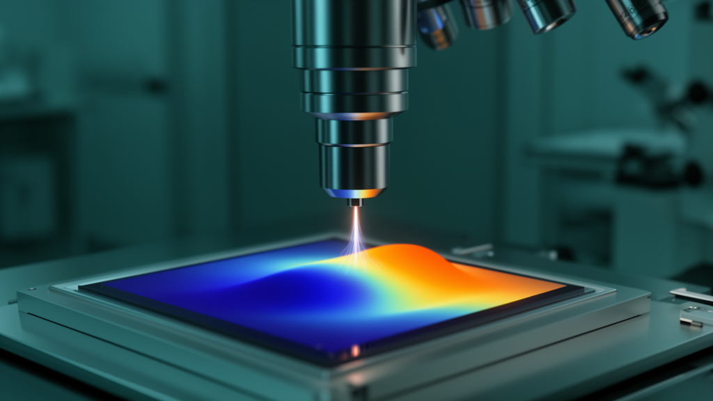
Measuring the bandgap of a material sounds like a solved problem: shine light on it, watch what it absorbs, and read off the answer. But as electronic devices shrink toward the atomic scale, the bandgap stops being a single number. It varies dramatically across defects, interfaces, strain fields, and quantum-confined structures, and conventional optical spectroscopy averages over all of it. A new review published in Applied Microscopy surveys how monochromated scanning transmission electron microscopy combined with valence electron energy-loss spectroscopy, or STEM-VEELS, has matured from a qualitative technique into a candidate platform for quantitative nanoscale bandgap metrology.
The principle is elegant. In STEM-VEELS, a focused beam of fast electrons passes through an ultrathin specimen, and the energy lost by electrons that excite valence electrons is recorded. Below the bandgap energy, transitions from the valence band to the conduction band are forbidden, so the interband signal is essentially absent. Once the energy loss exceeds the gap, allowed transitions appear as a characteristic onset in the spectrum. Because the electron probe can be focused to sub-nanometer dimensions, the technique can, in principle, locate that onset at a single point inside a quantum dot, across a buried interface, or within a single moiré unit cell.
The decisive hardware advance was the electron monochromator. Conventional cold field-emission guns deliver an energy spread of roughly 300 millielectronvolts, which blurs the weak interband signal into the tail of the intense zero-loss peak, the pile-up of unscattered and elastically scattered electrons at zero energy loss. Wien-filter and Omega-type monochromators now narrow the spread to below 50 to 120 millielectronvolts. The review highlights a demonstration at 80 kilovolts achieving an energy resolution of about 0.12 electronvolts, defined as the full width at half maximum of the zero-loss peak, sufficient to resolve subtle onset variations in narrow-gap semiconductors.
Extracting a reliable number, however, is far harder than detecting an edge. The zero-loss peak’s tail can exceed the intrinsic onset signal by orders of magnitude, so rigorous subtraction is the single most critical processing step. Researchers use vacuum reference spectra, dual-EELS acquisition, mirrored-tail estimates, and analytical fits based on Gaussian, Lorentzian, pseudo-Voigt, or Pearson functions. Each choice carries methodological bias, and different onset criteria applied to the same spectrum can yield different bandgap values. The review stresses that the reported bandgap is therefore not purely a material property but also a product of the chosen analysis workflow, making transparent reporting and uncertainty evaluation essential.
Physics itself imposes a further limit. Unlike core-loss excitations, low-loss valence excitations are governed by dipole selection rules and are strongly delocalized. The characteristic delocalization length scales roughly with the incident beam energy divided by the energy loss, so for typical semiconductor gaps of one to three electronvolts, the interaction volume can extend over several to tens of nanometers even with an atomic-scale probe. The effective spatial resolution of a bandgap measurement is therefore substantially poorer than the nominal probe size, and the review warns explicitly against equating probe diameter with measurement resolution.
Parasitic electromagnetic excitations add another layer of difficulty. When the electron velocity exceeds the phase velocity of light in the specimen, coherent Cherenkov radiation is emitted, injecting extra intensity into exactly the low-loss region where bandgap onsets appear. High-refractive-index, narrow-gap materials such as silicon, germanium, gallium arsenide, and indium phosphide are especially susceptible at conventional accelerating voltages of 200 to 300 kilovolts. Guided optical modes and other retardation-related effects contribute similar artifacts. Mitigation strategies include lowering the accelerating voltage, thinning specimens, and, most effectively, off-axis and Bessel-aperture acquisition geometries that exclude low-momentum-transfer electrons during illumination and collection.
The payoff of these advances is visible in the applications the review compiles. In individual lead sulfide quantum dots, monochromated STEM-VEELS measured a local bandgap of about 1.1 electronvolts, far above the bulk value of 0.41 electronvolts, with the widening attributable to quantum confinement, and mapped systematic variations from dot center to edge. In a multilayer gallium nitride heterostructure, off-axis Bessel STEM-EELS resolved distinct local bandgaps of approximately 3.3, 5.0, and 5.6 electronvolts for GaN, AlGaN, and AlN layers across a buried interface. At a beta-phase aluminum gallium oxide and gallium oxide heterointerface, the technique detected a localized bandgap dip of roughly 0.25 electronvolts attributed to interface-related defect states after correcting for beam delocalization.
Low-dimensional materials showcase the technique’s full spatial power. In twisted molybdenum disulfide and tungsten diselenide van der Waals bilayers, spatially resolved low-loss spectra revealed stacking-dependent electronic excitations within individual moiré supercells. In one-dimensional molybdenum disulfide nanotubes, combined analysis of excitonic features near 2 to 3 electronvolts, a plasmon resonance near 8.3 electronvolts, and bandgap maps showed that highly curved end regions exhibit a measurable bandgap reduction linked to curvature-induced tensile strain and structural defects. Machine-learning frameworks such as EELSfitter, which treat zero-loss peak subtraction as a statistical inference problem using neural networks and Monte Carlo sampling, made these uncertainty-aware maps possible, and have been extended to automated mapping of thickness, bandgap, and dielectric function in two-dimensional materials such as InSe flakes.
The review also looks forward. Cryogenic operation is expected to suppress beam damage and thermal broadening in fragile perovskites and van der Waals materials. Ultrafast pump-probe schemes and operando biasing promise real-time observation of transient electronic dynamics during device operation. Artificial intelligence is anticipated to automate hyperspectral analysis, although the authors caution that fully autonomous quantitative bandgap metrology has not yet been established. In parallel, a monochromated scanning electron microscope based reflection electron energy-loss spectroscopy system, achieving roughly 50 millielectronvolts energy resolution, was recently demonstrated on bulk specimens including graphene, graphite, gold, and silicon dioxide, resolving a bandgap onset near 9 electronvolts in silicon dioxide without observable Cherenkov contributions, offering a nondestructive complement for surfaces that cannot be thinned for transmission work.
Perhaps the review’s central message is that instrument performance is no longer the bottleneck. Modern systems routinely deliver sub-100 millielectronvolt energy resolution and nanometer-scale spatial resolution, so the pressing question is whether measured values are reliable, reproducible, and comparable across laboratories. The authors argue that standardized protocols for specimen preparation, acquisition, zero-loss peak treatment, onset determination, and uncertainty quantification are what will transform local bandgap measurement from a characterization capability into a traceable metrological technique, one that could underpin the design and quality assessment of next-generation semiconductor, quantum, and energy materials.
Subject of Research: A review of monochromated STEM-VEELS for nanoscale local bandgap measurement
Article Title: Local bandgap and optoelectronic measurement using monochromated STEM-VEELS: fundamentals, challenges, and recent advances
Article References: Jung, H. J. (2026). Local bandgap and optoelectronic measurement using monochromated STEM-VEELS: fundamentals, challenges, and recent advances. Applied Microscopy, 56(1), Article 22. https://doi.org/10.1186/s42649-026-00143-9
Image Credits: AI Generated
DOI: 10.1186/s42649-026-00143-9
Keywords: STEM-VEELS, bandgap, electron energy-loss spectroscopy, monochromator, zero-loss peak, Cherenkov radiation, quantum dots, two-dimensional materials, machine learning, spatial delocalization, SEM-REELS, nanoscale metrology
Cite Scienmag News
Denise Maddox. (October 28, 2026). Electron microscope technique maps bandgaps at the nanometer scale. Scienmag. https://scienmag.com/electron-microscope-technique-maps-bandgaps-at-the-nanometer-scale/
Denise Maddox. "Electron microscope technique maps bandgaps at the nanometer scale." Scienmag, 28 October 2026, https://scienmag.com/electron-microscope-technique-maps-bandgaps-at-the-nanometer-scale/. Accessed 28 October 2026.
Denise Maddox. "Electron microscope technique maps bandgaps at the nanometer scale." Scienmag. October 28, 2026. https://scienmag.com/electron-microscope-technique-maps-bandgaps-at-the-nanometer-scale/