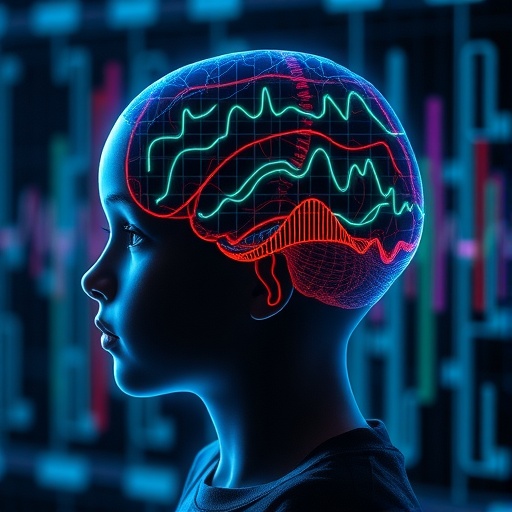
In a groundbreaking exploration that delves deep into the neural underpinnings of early childhood motor development, a recent systematic review has shed unprecedented light on the intricate EEG (electroencephalogram) signatures associated with this critical phase of human growth. As motor skills lay the foundation for a child’s interaction with the environment, understanding their cerebral correlates holds transformative potential for both pediatric neuroscience and clinical interventions targeting developmental disorders.
The study, meticulously conducted by Khurana, Kulkarni, Bulea, and colleagues, examines a compendium of EEG data spanning numerous longitudinal and cross-sectional studies. Their systematic approach elucidates patterns of neural oscillatory dynamics that mirror the maturation of motor circuits. By analyzing spectral power changes, coherence, and event-related potentials (ERPs), the review charts a comprehensive map of neurophysiological alterations that accompany the gradual refinement of motor proficiency in infants and toddlers.
One of the pivotal revelations of the research lies in identifying specific frequency bands that evolve in their prominence and interaction as motor development progresses. Notably, the mu rhythm (8-13 Hz), traditionally linked to sensorimotor processes, demonstrates a characteristic trajectory where its desynchronization intensifies with advancing motor abilities. This finding substantiates the hypothesis that mu rhythm modulation serves as a robust biomarker for integrity and maturation of the sensorimotor cortex during early childhood.
Furthermore, the systematic review highlights the dynamic interplay between beta oscillations (13-30 Hz) and motor activity. Beta rhythms, often implicated in motor control and inhibition, show distinctive event-related desynchronization during active movement phases in young children. Intriguingly, the authors emphasize that the amplitude and temporal dynamics of beta oscillations provide critical insight into the neurodevelopmental timeline, potentially offering early signals of atypical motor development when deviations from normative EEG patterns are observed.
Beyond oscillatory activity, coherence measures detailed in the review illuminate how the functional connectivity between cortical motor regions evolves. The strengthening of interhemispheric synchronization correlates with milestones such as grasping, crawling, and walking, indicating a neural integration process that underpins increasingly complex motor coordination. These connectivity signatures not only reflect anatomical maturation but also the refinement of sensorimotor loops essential for skilled movements.
The review also explores the role of event-related potentials (ERPs), demonstrating how specific components linked to motor planning and execution emerge and shift as children transition from reflexive to volitional movements. The N200 and P300 components, noteworthy for their association with attentional and cognitive control aspects of motor function, exhibit maturation patterns consistent with enhanced motor control and learning capacity. This dual emphasis on oscillatory activity and ERPs furnishes a multidimensional understanding of how the developing brain orchestrates motor behavior.
Intriguingly, the authors discuss the impact of environmental and experiential factors on EEG signatures. Infants exposed to enriched motor experiences, such as interactive play and motor skill training, tend to exhibit expedited development of sensorimotor rhythms and connectivity. This observation underscores the neuroplastic potential inherent in early childhood and advocates for intervention strategies that leverage activity-dependent brain remodeling to optimize motor outcomes, especially in at-risk populations.
A significant portion of the review addresses methodological considerations pertinent to pediatric EEG research. The inherent challenges of recording reliable EEG data in young children, including movement artifacts and compliance issues, are scrutinized. The authors emphasize advanced computational approaches to artifact mitigation and the importance of harmonizing protocols across studies to build robust, comparable datasets. Their recommendations pave the way for future research to harness high-density EEG and machine learning for predictive modeling in developmental neuroscience.
Clinically, this systematic review charts promising avenues for early diagnosis and intervention in neurodevelopmental disorders characterized by motor deficits, such as cerebral palsy and autism spectrum disorder. By establishing normative EEG trajectories, deviations can be detected with greater sensitivity, facilitating timely therapeutic measures. The integration of EEG biomarkers with behavioral assessments offers a comprehensive framework for personalized medicine approaches in pediatric neurology.
Moreover, the longitudinal perspective adopted in the review captures the non-linear and individualized nature of motor development. The authors acknowledge that while overarching EEG trends exist, substantial inter-individual variability is noted, reflecting genetic, environmental, and experiential heterogeneity. This nuanced understanding cautions against oversimplified interpretations and calls for personalized monitoring frameworks that accommodate diverse developmental trajectories.
On a technological frontier, the review spotlights emerging neuroimaging modalities synergistic with EEG, such as functional near-infrared spectroscopy (fNIRS), which can complement electrophysiological data with hemodynamic insights. Such multimodal approaches can unravel complex neurovascular and neuroelectrical coupling during motor tasks, offering a richer depiction of early brain function.
Importantly, the review calls attention to the translational gap that persists between laboratory findings and community applications. Developing scalable, child-friendly EEG assessment tools that can be deployed in pediatric clinics, schools, and even homes is identified as a critical future goal. Enhanced portability and user-interface designs will democratize access to objective neurophysiological metrics of motor development.
In summarizing, the comprehensive survey of EEG evidence presented collectively affirms that early childhood motor development is characterized by distinct, quantifiable changes across multiple neurophysiological dimensions. From rhythmic oscillations reflecting sensorimotor engagement to connectivity patterns signaling cortical maturation, the brain’s electrical dynamics offer a powerful window into developmental progress.
This research not only advances theoretical understanding within developmental neuroscience but also propels the field toward innovative diagnostics and tailored interventions. As motor development constitutes a keystone for broader cognitive and social capacities, elucidating its neural signatures empowers caregivers, clinicians, and scientists alike to foster optimal outcomes during this formative window of human life.
With motley advances in data analytics and wearable technology, the horizon for EEG-based monitoring is bright. The convergence of neurobiological insights with pragmatic tools heralds a future where early detection of motor impairments is routine, enabling preemptive measures that significantly enhance children’s quality of life.
This systematic review by Khurana et al. thus marks a seminal contribution to pediatric neuroscience, encapsulating a rich synthesis of current knowledge and shaping the trajectory for future research. The evolving landscape promises that unraveling the brain’s motor signatures will continue to illuminate the profound complexities of early childhood development, catalyzing breakthroughs that resonate across science and society.
Subject of Research: EEG Signatures of Motor Development in Early Childhood
Article Title: EEG signatures of motor development in early childhood: a systematic review
Article References:
Khurana, S., Kulkarni, A., Bulea, T.C. et al. EEG signatures of motor development in early childhood: a systematic review. Pediatr Res (2026). https://doi.org/10.1038/s41390-026-05160-8
Image Credits: AI Generated
DOI: 13 June 2026
