


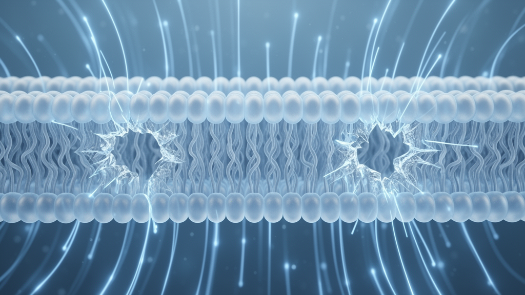
Nanosecond pulsed electric fields have emerged as a promising non-thermal modality for tumor treatment, offering a method to induce irreversible cellular damage without the thermal side effects associated with conventional ablation techniques. Central to this process is electroporation, a phenomenon where external electric fields increase the permeability of biological membranes, leading to the formation of nanoscale pores. While the plasma membrane, which encloses the cell, has been the primary focus of electroporation research, recent attention has shifted toward the nuclear envelope, the double-membrane structure that houses genetic material. Understanding how these distinct membranes respond to electric fields is critical for optimizing cancer therapies, as the integrity of the nuclear envelope is closely linked to tumor initiation and progression.
A new study published in PLoS ONE by researchers from the University of Shanghai for Science and Technology provides a molecular-level explanation for why the nuclear envelope may be more susceptible to damage than the plasma membrane under nanosecond pulsed electric fields. Using molecular dynamics simulations, the team compared the electroporation responses of a single phospholipid bilayer, representing the plasma membrane, and a double phospholipid bilayer, representing the nuclear envelope, under identical electric field strengths. The findings reveal that the double membrane undergoes faster electroporation due to a unique mechanical interaction between its two layers, a process that significantly reduces membrane tension and facilitates pore formation.
The study employed coarse-grained molecular dynamics simulations to model the membranes, utilizing a Martini force field to represent the lipid components. The researchers constructed a single membrane model composed of 1,024 dipalmitoylphosphatidylcholine molecules and a double membrane model consisting of two such bilayers separated by a 21.6 nanometer intermembrane space filled with a 0.15 M sodium chloride solution. This setup allowed for a controlled comparison of the fundamental structural difference between the two membrane types: the number of phospholipid bilayers. By applying constant electric fields ranging from 0.2 to 0.6 volts per nanometer, the simulations captured the rapid dynamics of pore formation on nanosecond timescales, a regime that is difficult to observe experimentally.
The results demonstrated a clear disparity in electroporation kinetics between the two systems. Under the same electric field conditions, the double membrane consistently required less time to form pores than the single membrane. As the electric field strength increased from 0.2 to 0.6 volts per nanometer, the pore formation time for the double membrane decreased from 0.6 to 0.2 nanoseconds, whereas the single membrane’s formation time decreased from 4.6 to 0.3 nanoseconds. Statistical analysis confirmed that these differences were highly significant, with a p-value of less than 0.0001. This suggests that the nuclear envelope is more susceptible to electroporation-induced damage than the plasma membrane, a finding that aligns with previous cellular-scale observations of greater structural damage to intracellular organelles.
To understand the mechanism behind this accelerated pore formation, the researchers analyzed the forces acting on the membranes. They found that the double membrane experienced lower Coulomb force in the plane perpendicular to the membrane normal, while exhibiting higher Coulomb force along the direction parallel to the membrane normal compared to the single membrane. This redistribution of forces is driven by a phenomenon known as inter-membrane counter-deformation. Under the influence of the electric field, the upper and lower bilayers of the double membrane deform in opposite directions, creating local regions where the two layers approach each other. This counter-deformation alters the local electric field angle distribution, shifting the membrane normal toward smaller angles relative to the field direction.
This geometric shift has profound implications for membrane tension. The study showed that the reduction in membrane tension was markedly greater in the double membrane than in the single membrane. At an electric field strength of 0.2 volts per nanometer, the membrane tension in the double membrane decreased by approximately 106.5 millinewtons per meter, compared to a reduction of only 10.9 millinewtons per meter in the single membrane. At higher field strengths, the disparity widened further, with the double membrane experiencing a tension drop of 741.0 millinewtons per meter versus 277.4 millinewtons per meter for the single membrane. The analysis indicated that this significant tension reduction lowers the energy barrier for pore formation, thereby accelerating the electroporation process in the double membrane system.
Further characterization of the pores revealed that the double membrane not only formed pores faster but also exhibited a greater number and larger size of pores compared to the single membrane. The pore area ratio, quantified as the fraction of the membrane surface unoccupied by lipid atoms, was consistently higher in the double membrane across various time points and field strengths. For instance, at 0.6 nanoseconds and an electric field of 0.3 volts per nanometer, the pore area percentage was 12.60% for the double membrane, while it was only 0.06% for the single membrane. Over longer timescales, multiple adjacent pores in the double membrane were observed to expand and coalesce, forming interconnected channels that enable bidirectional solution transport, potentially leading to more severe cellular damage.
The researchers also investigated the role of lipid headgroup orientation in this process. They found that as the electric field strength increased, the lipid headgroups in both membrane systems reoriented to align more closely with the electric field direction. In the single membrane, the peak of the headgroup angle distribution shifted from 75 degrees to 30 degrees as the field increased from 0.2 to 0.6 volts per nanometer. This reorientation contributes to the increased Coulomb force component parallel to the membrane normal and the reduced perpendicular component, further facilitating pore formation. However, the counter-deformation specific to the double membrane amplifies these effects, leading to a more pronounced shift in the electric field angle distribution and a more substantial reduction in membrane tension.
Despite the insights provided by this study, the authors acknowledge several limitations. The simulations used a coarse-grained pure dipalmitoylphosphatidylcholine model, which neglects key features of native membranes such as compositional asymmetry, cholesterol content, and the presence of membrane proteins. These factors are known to influence electroporation behavior and membrane mechanics. Additionally, the study focused on the mechanical and electrostatic aspects of electroporation without considering thermal effects, although previous research has shown that temperature can enhance electroporation by increasing electrical conductivity and reducing membrane tension. Future work will need to incorporate more complex, cell-type-specific membrane models to fully understand the implications of these findings for clinical applications.
This research provides a molecular-scale theoretical reference for understanding the differential effects of nanosecond pulsed electric fields on the plasma membrane and the nuclear envelope. By elucidating the role of inter-membrane counter-deformation in accelerating electroporation, the study highlights the nuclear envelope as a potential target for enhancing the efficacy of cancer treatments. As cancer cells often exhibit enlarged nuclei and increased nucleocytoplasmic ratios, targeting the nuclear envelope could offer a novel strategy for inducing irreversible cell damage. The findings underscore the importance of considering intracellular structures in the design of electroporation therapies, moving beyond the traditional focus on the plasma membrane to achieve more effective tumor ablation.
Subject of Research: Molecular dynamics simulation of electroporation kinetics and membrane tension in single versus double phospholipid bilayers under nanosecond pulsed electric fields.
Article Title: Tension reduction from inter-membrane counter-deformation accelerates nuclear electroporation
Article References: Diao, Y., Ye, P., & Liu, B. (2026). Tension reduction from inter-membrane counter-deformation accelerates nuclear electroporation. PLOS One, 21(10), e0361048. https://doi.org/10.1371/journal.pone.0361048
Image Credits: AI Generated
DOI: 10.1371/journal.pone.0361048
Keywords: electroporation, nanosecond pulsed electric fields, nuclear envelope, plasma membrane, molecular dynamics, membrane tension, counter-deformation, cancer treatment, lipid bilayer, pore formation, Tension, reduction
Cite Scienmag News
Nathaniel Bowman. (October 23, 2026). Double Membranes Puncture Faster Under Electric Fields. Scienmag. https://scienmag.com/double-membranes-puncture-faster-under-electric-fields/
Nathaniel Bowman. "Double Membranes Puncture Faster Under Electric Fields." Scienmag, 23 October 2026, https://scienmag.com/double-membranes-puncture-faster-under-electric-fields/. Accessed 23 October 2026.
Nathaniel Bowman. "Double Membranes Puncture Faster Under Electric Fields." Scienmag. October 23, 2026. https://scienmag.com/double-membranes-puncture-faster-under-electric-fields/


