


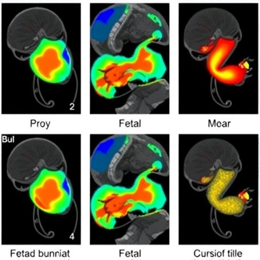
Developmental venous anomalies, the most common cerebral vascular malformations, were detected in 0.46 percent of fetuses undergoing magnetic resonance imaging at a large tertiary center, according to a retrospective study that also identified two reproducible imaging patterns allowing prenatal diagnosis without contrast agents or susceptibility-weighted imaging. The findings, drawn from nearly 4,000 fetal MRI examinations performed between January 2017 and June 2025, suggest that these congenital venous malformations can be recognized in utero with far greater confidence than previously appreciated, potentially transforming prenatal counseling for families whose fetuses show unexplained susceptibility artifacts or echogenic lesions on ultrasound.
The research, led by Stephanie Libzon and Liat Ben Sira of Tel Aviv Sourasky University Medical Center and published in Pediatric Radiology, systematically reviewed the fetal MRI database at their institution. Of 3,932 fetal MRIs, 195 examinations (about 5 percent) had shown artifacts on hemorrhage-sensitive sequences in the original reports. The research team extracted cases with a documented developmental venous anomaly diagnosis and, crucially, re-evaluated every study with susceptibility artifacts suggestive of hemorrhage, vascular malformation, or even questionable hemosiderin, to catch anomalies that had not been recognized initially. Eighteen fetuses were ultimately identified with developmental venous anomalies, corresponding to the observed detection rate of 0.46 percent. Two of these cases were found only upon the retrospective review, underscoring how easily such lesions can be missed without a systematic search.
Two independent reviewers — a pediatric neuroradiologist with 25 years of experience and a PhD student with expertise in fetal neuroradiology — assessed each case while blinded to the other’s assessments. Inter-rater agreement was excellent, with a Cohen’s kappa of 0.83 (95 percent confidence interval, 0.69–0.96), indicating near-perfect concordance despite the rarity of positive cases. When the two reviewers disagreed, interpretations were reconciled through consensus discussions with a third pediatric radiologist with 30 years of experience in fetal medicine. The reviewers confirmed the diagnosis and evaluated the number of lesions, their location, their drainage direction — superficial, deep, or mixed — and associated abnormalities such as draining venous varix thrombosis, adjacent parenchymal hemorrhage, polymicrogyria, or restricted diffusion. Fetuses with any such findings, multiple anomalies, or parenchymal loss at the anomaly’s insertion site were classified as having complicated developmental venous anomalies.
The demographic profile of the affected fetuses reflected typical patterns seen in postnatal series. The median maternal age was 32 years, the median gestational age at MRI was 33 weeks, and of the 17 fetuses with available sex data, 13 (about 77 percent) were male. More than half of the affected fetuses (10 of 18) had been referred for MRI because of echogenicity on prenatal ultrasound suspicious for hemorrhage or venous anomaly, while the remainder were referred for unrelated reasons including ventriculomegaly, periventricular pseudo-cysts, facial dysmorphism, and intrauterine growth restriction. Six cases were incidental findings noted at the time of the initial report, and the authors emphasize that these incidental detections highlight the importance of scrutinizing venous structures even when MRI is performed for other indications.
Anatomically, the anomalies showed a striking pattern. Frontal locations predominated, accounting for 10 of 18 cases (55.6 percent), followed by parieto-occipital (7 cases, 38.9 percent) and a single parietal lesion. Most lesions — 14 of 18, or 77.8 percent — were right-sided, a laterality consistent with prior postnatal reports, although the biological significance of this predominance remains unclear. Deep drainage was the most common drainage pathway (8 cases, 44.4 percent), with purely superficial drainage in 4 cases (22.2 percent) and mixed drainage in 6 (33.3 percent). All parieto-occipital lesions involved deep drainage. No infratentorial anomalies were found, which the authors attribute both to the genuine rarity of posterior fossa developmental venous anomalies and to the technical difficulty of detecting susceptibility-related changes in the posterior fossa on fetal MRI.
The heart of the study lies in the two characteristic imaging patterns the team identified. Because fetal MRI cannot use intravenous contrast and, in most cases, lacked dedicated susceptibility-weighted imaging, the researchers relied on gradient echo-based T2-weighted sequences — acquired routinely since 2017 — and, in half of the fetuses, T2* echo-planar imaging. The classic “caput medusae” configuration of radially oriented medullary veins was not observed prenatally in any case. Instead, on T2-weighted imaging, frontal anomalies with superficial drainage frequently showed what the authors call the “fisherman’s rope” sign: a collecting vein transversing the adjacent extra-axial fluid, accompanied by small linear hypointense lesions in the surrounding parenchyma, resembling a Japanese fisherman’s rope. This sign was significantly more common in fetuses with enlarged extra-axial spaces, seen in 5 of 6 such cases compared with 3 of 12 without (P=0.04), suggesting that generous fluid spaces make the superficial vein easier to trace.
The second pattern involves deep-draining anomalies, which appeared as small, focal, hypointense T2 irregular thickening of the ventricular wall. On coronal images, these lesions presented as hypointense areas that hug or envelop the superior margin of the lateral ventricle at the atrium level, without associated parenchymal loss or ventricular wall irregularity. This ventricular-wall hypointensity pattern has not previously been described for developmental venous anomalies and, critically, must be differentiated from hemorrhage, since the prognostic and counseling implications of the two entities differ substantially. Contrary to prior assumptions that these anomalies cannot be visualized on conventional T2-weighted sequences, the study demonstrated that high-resolution, thin-slice imaging — as thin as 2.5 millimeters in parasagittal planes — consistently depicted the superficial draining vein and, occasionally, subtle white matter hypointensities, usually without clear corresponding changes on T1-weighted imaging.
Eight fetuses (44.4 percent) had complicated anomalies, defined by the presence of parenchymal loss at the anomaly insertion site (6 cases), multiple anomalies (3 cases), draining venous varix thrombosis (2 cases), or adjacent parenchymal hemorrhage (2 cases). A notable statistical association emerged: thrombosis of a draining venous varix occurred exclusively in fetuses with multiple developmental venous anomalies (2 of 3 versus 0 of 15, P=0.02). The authors suggest this identifies a subgroup potentially at risk for vascular complications that may warrant closer prenatal and postnatal surveillance. Frontal anomalies with superficial drainage were also frequently associated with focal parenchymal loss at the venous insertion site — 3 of 4 purely superficial cases versus 3 of 14 others — an association that, while not statistically significant, suggests a topographic or hemodynamic relationship. Notably, no anomalies in this cohort were associated with cavernous malformations, which have been described in other pediatric and adult populations.
Beyond the anomalies themselves, five fetuses had concurrent ventriculomegaly, contralateral to the anomaly in three cases, ipsilateral in one, and bilateral and severe in one. Seven fetuses showed asymmetry of the medullary veins on susceptibility-sensitive sequences, and in six of these seven the asymmetry was on the side opposite the anomaly. The authors speculate that this may reflect venous flow redistribution, in which impaired outflow through the anomaly is compensated by contralateral venous dilatation, potentially raising venous pressures and contributing to ventricular enlargement — a mechanism supported by postnatal hemodynamic studies of symptomatic anomalies, though they caution that this remains speculative in the fetal setting. Additional findings included enlarged head biometry in five fetuses, small biometry in one, and enlarged extra-axial fluid spaces in six.
Follow-up data provided important validation. All 18 fetuses were liveborn, and four underwent more than one fetal MRI, confirming stability of the anomalies over time. Seven neonates underwent postnatal ultrasound and five underwent postnatal brain MRI at a median age of 12 days. All five postnatal MRIs confirmed the stable appearance of the prenatal findings, and all demonstrated the classical caput medusae appearance on susceptibility-weighted imaging that had not been visible on fetal MRI — a discrepancy the authors attribute to the limited sensitivity of fetal gradient echo sequences compared with postnatal susceptibility-weighted imaging. In all but one case, the anomalies remained stable in configuration and number; the exception was a neonate who had two anomalies and varix thrombosis prenatally and was found to have multiple anomalies postnatally without parenchymal hemorrhage, and remains under investigation.
A major practical contribution of the study is its framework for distinguishing developmental venous anomalies from periventricular venous hemorrhagic infarction, a critical distinction given their different pathophysiology and prognosis. On thin-section T2-weighted imaging, hemorrhagic infarction typically shows marked parenchymal hypointensity reflecting blood products, whereas venous anomalies often appear nearly normal with only subtle focal hypointensities. On coronal images, hemorrhagic infarction forms an upward triangular configuration of engorged medullary veins extending toward the ventricular margin, while deep anomalies display a downward triangular pattern hugging the ventricle’s superior margin. Parenchymal injury also differs: in infarction it abuts the ventricle, while in anomalies it tends to be remote, at the insertion site of a superficial draining vein. Perhaps most tellingly, anomalies are stable over time, whereas hemorrhagic lesions evolve according to the age of the bleed, sometimes culminating in porencephalic cysts. The discordance between a hyperechogenic lesion on ultrasound and delicate, non-evolving hypointensity on MRI provides a key diagnostic indicator.
The authors acknowledge important limitations. The study was retrospective, conducted at a single tertiary hospital, and included a relatively small number of cases; referral of fetuses for abnormal brain ultrasound may have introduced selection bias. The absence of contrast-enhanced sequences could have led to under-recognition of subtle anomalies, and not all cases had confirmatory postnatal imaging. The observed fetal prevalence of 0.46 percent is lower than reported neonatal rates of 0.8 to 1.9 percent and far below the 5 to 10 percent seen in older children and adults, likely reflecting a combination of technical limitations — motion artifacts, limited spatial resolution, no contrast — and the possibility that anomaly prevalence genuinely increases with age. One excluded case of cerebellar susceptibility artifacts initially interpreted as capillary telangiectasia illustrates the ongoing challenge of distinguishing venous anomalies from other vascular malformations prenatally.
Nevertheless, the implications for clinical practice are considerable. The study confirms that hemorrhage-sensitive gradient echo-based T2-weighted and T2* echo-planar sequences provide sufficient diagnostic information in the prenatal setting, supporting their routine inclusion in fetal MRI protocols. Combined with high-resolution thin-slice T2-weighted imaging, coronal planes, and serial imaging to document stability, these sequences enable confident prenatal diagnosis without the postnatal “gold standard” of contrast-enhanced imaging. For families facing the discovery of a fetal brain lesion, the ability to distinguish a largely benign, congenital venous anomaly from a hemorrhagic infarction or other vascular malformation could meaningfully alter counseling, surveillance, and management — and, as the authors conclude, supports a more standardized approach to fetal MRI assessment of the cerebral venous system.
Cite Scienmag News
Colin Clarke. (August 31, 2026). Developmental venous anomalies on fetal magnetic resonance imaging: prevalence and reproducible radiological phenotypes. Scienmag. https://scienmag.com/developmental-venous-anomalies-on-fetal-magnetic-resonance-imaging-prevalence-and-reproducible-radiological-phenotypes/
Colin Clarke. "Developmental venous anomalies on fetal magnetic resonance imaging: prevalence and reproducible radiological phenotypes." Scienmag, 31 August 2026, https://scienmag.com/developmental-venous-anomalies-on-fetal-magnetic-resonance-imaging-prevalence-and-reproducible-radiological-phenotypes/. Accessed 3 September 2026.
Colin Clarke. "Developmental venous anomalies on fetal magnetic resonance imaging: prevalence and reproducible radiological phenotypes." Scienmag. August 31, 2026. https://scienmag.com/developmental-venous-anomalies-on-fetal-magnetic-resonance-imaging-prevalence-and-reproducible-radiological-phenotypes/


