


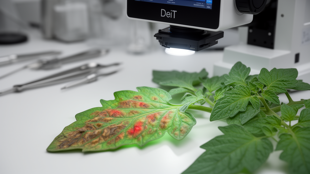
A hybrid artificial-intelligence system achieved 99.13% accuracy in classifying 38 healthy and diseased plant-leaf categories while producing visual explanations designed to show both localized lesions and broader image context. The study, published as open-access research, evaluated Data-efficient Image Transformers (DeiT) on the augmented New Plant Diseases Dataset, which contains 70,295 training images and 17,572 validation images spanning 14 plant species. Its strongest model, DeiT-Base, also received a 17.00-millisecond average inference time for a 224 × 224 RGB image on an NVIDIA Tesla T4 GPU, according to the authors. These results apply to a controlled benchmark dataset and do not establish equivalent performance in field conditions.
The framework was designed to address two weaknesses commonly associated with image-based plant disease models: limited transparency and the inability of many systems to estimate how extensively a leaf is affected rather than simply naming a disease class. Researchers Muskan Batra and Pooja Sharma combined three DeiT variants—Tiny, Small and Base—with an Explainable Artificial Intelligence module and a post-classification severity estimator. The classifier works on the original RGB image, while a separate color-based segmentation stage is applied only after prediction to isolate the leaf and estimate visible damage. This distinction is important because the segmentation module does not improve classification accuracy; it supports explanation refinement and severity estimation.
At the technical center of the classifier is the transformer’s self-attention mechanism, which allows image patches to interact across the entire frame rather than relying only on local convolutional filters. Each 224 × 224 image was divided into non-overlapping 16 × 16-pixel patches, and the patches were linearly projected into embeddings. Positional embeddings preserved their spatial relationships, while a special classification token passed through stacked encoder layers and into a fully connected classification head. The three models began with ImageNet-pretrained weights. During fine-tuning, the backbone was frozen and only the classification head was optimized with AdamW, cross-entropy loss and a learning rate of 5 × 10⁻⁴. Training used random horizontal flipping, rotations of plus or minus 15 degrees, ImageNet normalization and mixed-precision computation.
DeiT-Base performed best among the tested transformers and convolutional baselines. Its reported accuracy was 99.13%, compared with 98.65% for DenseNet121, the strongest CNN baseline. MobileNetV3 reached 97.63%, while ResNet50, EfficientNet-B4 and InceptionV3 also underwent training and evaluation under the same data split and experimental conditions. Three independent DeiT-Base runs using random seeds 42, 123 and 999 produced an average accuracy of 99.1167% with a standard deviation of 0.0404 percentage points. Average training time was 134.54 minutes with a standard deviation of 0.94 minutes per run. The normalized confusion matrix was strongly diagonal, although some confusion remained among diseases with similar appearances, including tomato early blight, late blight and target spot.
The researchers paired Grad-CAM with Attention Rollout to produce a unified local-global explanation. Grad-CAM uses gradients associated with the predicted class to identify spatially influential features, such as lesions, spots and discolored tissue. Because DeiT lacks conventional convolutional feature maps, the method was applied to the model’s final feature representations before the classification head. Attention Rollout instead aggregates attention across transformer layers, revealing how patches influence one another over long spatial distances. The two normalized maps were fused with the hybrid equation H = 0.6HGrad-CAM + 0.4HAttention Rollout, after which normalization, Gaussian smoothing and thresholding reduced scattered or low-activation regions.
Across all 38 classes, the hybrid map achieved a Focus Score of 0.9771 and Background Noise of 0.0229, where higher focus and lower background noise are considered preferable. Its signal-to-noise ratio was 61.1871 and its entropy was 9.9726; lower entropy indicates a more concentrated spatial distribution. Grad-CAM recorded the highest Faithfulness score, 0.7917, meaning that masking the input with its highlighted regions preserved the target-class probability more strongly than the hybrid method. The researchers interpreted this as a trade-off: Grad-CAM offered stronger prediction-preserving localization, while Attention Rollout supplied broader contextual information. An ablation study found that a fusion weight of 0.8 produced slightly better localization metrics, but the authors retained 0.6 to preserve a larger contribution from global attention.
Severity estimation used two complementary ratios. The damage ratio was the number of pixels identified as infected by HSV thresholds divided by the total segmented leaf area. Brown and yellow regions were treated as potential disease symptoms, with Gaussian smoothing used to improve continuity. The XAI ratio was the area covered by the top 25% of hybrid-map activations divided by the leaf area. The final score was calculated as 100 × (0.6 × damage ratio + 0.4 × XAI ratio). Scores from 0% to 25% were labeled low, 25% to 60% moderate and above 60% severe. A separate adjustment rule raises a score above 60% when the visible damage ratio itself exceeds 50%, preventing extensive damage from being classified as less severe solely because attention is dispersed.
Representative outputs tracked visible symptoms: an apple-scab example received a 10.7% score, tomato target spot 9.0%, apple cedar rust 37.6%, corn cercospora leaf spot 29.6%, potato early blight 35.3% and tomato late blight 35.5%. Strawberry leaf scorch, a more extensively affected example, was scored at 61.3%. These observations are computational estimates rather than validated measurements of disease progression. The dataset contains no expert-annotated severity labels, so the researchers did not compare the scores with agronomist or plant-pathologist assessments. Likewise, the XAI evaluation used HSV-derived leaf masks as a proxy for relevant regions rather than expert-marked lesions. Its metrics therefore assess concentration within the segmented leaf and suppression of background activations, not whether a heatmap precisely matches pathological annotations.
Although the results are striking, the study’s scope limits what can be inferred. No independent test set, cross-dataset validation or cross-validation was performed, and the researchers noted that visually similar samples cannot be completely excluded from the official split. The augmented benchmark contains relatively controlled imagery, whereas real farms introduce changing illumination, shadows, occlusion, cluttered backgrounds, different camera angles and wider variation in disease appearance. HSV segmentation may also be sensitive to those conditions. The study also compared only the three DeiT sizes and five CNN baselines, not alternative transformer designs such as Swin Transformer, ConvNeXt, MobileViT, MaxViT or EfficientFormer, and it did not benchmark hybrid explanations against methods such as Score-CAM, Integrated Gradients, SHAP or LIME. With 85.83 million parameters and a reported 16.85 GFLOPs per image, DeiT-Base is not yet a lightweight field device.
The authors position the system as decision support rather than a replacement for expert diagnosis and note that false-negative classifications or inaccurate severity estimates could affect agricultural management. Their next steps include independent field evaluation, expert-annotated severity data, more robust segmentation, comparison with other vision transformers and explainability methods, and optimization for mobile or edge hardware. Even so, the study demonstrates a technically coherent attempt to move beyond a single disease label. It joins a high-performing image classifier with two kinds of visual evidence—local symptom attribution and global patch relationships—then exposes the combination as a numerical severity indicator. Whether that indicator proves useful in practice will depend on validation beyond the standardized benchmark.
Subject of Research: Explainable DeiT models for plant disease classification and severity estimation on a benchmark leaf-image dataset
Article Title: Hybrid explainable DeiT-based framework for plant disease classification and severity estimation
Article References: Batra, M., & Sharma, P. (2026). Hybrid explainable DeiT-based framework for plant disease classification and severity estimation. Discover Artificial Intelligence, 6(1), Article 1109. https://doi.org/10.1007/s44163-026-02191-2
Image Credits: AI Generated
DOI: 10.1007/s44163-026-02191-2
Keywords: plant disease detection, Data-efficient Image Transformer, explainable AI, Grad-CAM, Attention Rollout, disease severity estimation, precision agriculture, image segmentation, deep learning, plant pathology, Hybrid, explainable
Cite Scienmag News
Alan Morgan. (October 26, 2026). DeiT framework pairs plant disease detection with interpretable severity estimates. Scienmag. https://scienmag.com/deit-framework-pairs-plant-disease-detection-with-interpretable-severity-estimates/
Alan Morgan. "DeiT framework pairs plant disease detection with interpretable severity estimates." Scienmag, 26 October 2026, https://scienmag.com/deit-framework-pairs-plant-disease-detection-with-interpretable-severity-estimates/. Accessed 26 October 2026.
Alan Morgan. "DeiT framework pairs plant disease detection with interpretable severity estimates." Scienmag. October 26, 2026. https://scienmag.com/deit-framework-pairs-plant-disease-detection-with-interpretable-severity-estimates/


