


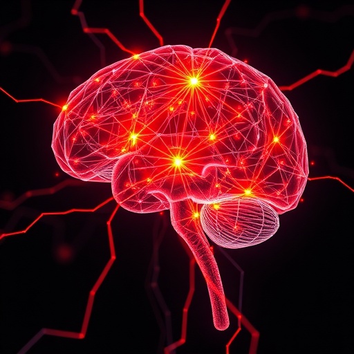
In an era where neurological disorders pose significant challenges to medical science, a groundbreaking study has emerged that may revolutionize the way we understand and diagnose Parkinson’s disease. Researchers led by Shin HG, Mills KA, and Dawson TM have unveiled a novel method for in-vivo iron mapping in patients with Parkinson’s disease, harnessing the power of deep learning-driven magnetic resonance imaging (MRI) techniques. The findings, published in the prestigious journal npj Parkinson’s Disease in 2026, promise not only to enhance our diagnostic capabilities but also to deepen our comprehension of the pathophysiological mechanisms underlying this debilitating condition.
Iron accumulation in specific brain regions has long been associated with neurodegenerative diseases, including Parkinson’s. Traditionally, detecting and quantifying iron in vivo within the brain posed considerable challenges due to the complex magnetic properties of iron and the limitations of conventional imaging technologies. However, the interdisciplinary team capitalized on the latest advancements in deep learning and MRI technology to address these challenges by developing what they call a susceptibility source separation technique. This approach innovatively separates signals arising from various magnetic sources, specifically isolating iron-related susceptibility changes in brain tissues.
The core of this method hinges on susceptibility-weighted imaging (SWI), a magnetic resonance technique sensitive to magnetic susceptibility variations caused by substances like iron, calcium, and blood products. While SWI has been used in neuroimaging before, its specificity for iron mapping was limited by confounding factors. To overcome this, the researchers integrated a deep neural network capable of disentangling these mixed signals. This system was trained on extensive datasets comprising both simulated and clinical MRIs, enabling it to learn intricate patterns and successfully isolate iron deposits with remarkable precision.
One of the most exciting implications of this method lies in its ability to conduct real-time in-vivo iron mapping, providing clinicians with unprecedented insight into the spatiotemporal distribution of iron accumulation across different brain regions. By visualizing these iron deposits, which are often concentrated in the substantia nigra and other basal ganglia structures implicated in Parkinson’s disease, researchers can better track disease progression and potentially identify early biomarkers that precede symptomatic onset.
The application of deep learning in this context exemplifies the transformative potential of artificial intelligence (AI) in medical imaging. By refining the separation of competing susceptibility signals, this technique transcends previous limitations that hindered quantification accuracy. Deep learning algorithms like the one employed in this study excel at recognizing subtle, nonlinear relationships within complex datasets that conventional analytical models fail to resolve. This capability significantly enhances the reliability of iron quantification, which is crucial for both research and clinical practice.
In addition to its diagnostic promise, in-vivo iron mapping through this deep learning-based susceptibility source separation MRI offers profound implications for therapeutic monitoring. As novel neuroprotective and disease-modifying treatments emerge, there is a critical need for reliable biomarkers to evaluate therapeutic effects objectively. By monitoring changes in brain iron concentrations longitudinally, clinicians and researchers can assess treatment efficacy more accurately and adjust strategies accordingly.
Parkinson’s disease remains a multifaceted disorder characterized by motor symptoms such as bradykinesia, rigidity, and resting tremor, alongside non-motor manifestations like cognitive decline and autonomic dysfunction. The condition’s etiology involves a complex interplay of genetic, environmental, and biochemical factors, among which iron accumulation has garnered significant attention. Excess iron is thought to catalyze oxidative stress and promote neuroinflammation, exacerbating neuronal loss in vulnerable regions. The ability to visualize and measure iron deposits noninvasively offers an invaluable window into these pathophysiological processes.
Technically, the researchers employed a multi-echo gradient echo sequence in their MRI protocol, facilitating the capture of phase images sensitive to magnetic susceptibility. These images provide raw data containing mixed susceptibility sources. The deep learning framework then processes this data, employing layers of convolutional neural networks to perform source separation. This approach enables extraction of distinct susceptibility maps corresponding specifically to iron distribution, eliminating interference from other magnetic substances.
The team validated their method on both animal models and human subjects diagnosed with Parkinson’s disease. The results demonstrated high correlation with post-mortem histological assessments, confirming the accuracy of iron mapping. Furthermore, the in-vivo maps revealed differential patterns of iron accumulation depending on disease severity and duration, suggesting that this technique could aid in disease staging and personalized medicine.
Beyond Parkinson’s, this approach has broader applicability for other neurodegenerative conditions involving abnormal iron metabolism, such as Alzheimer’s disease, multiple sclerosis, and Huntington’s disease. Iron dysregulation is increasingly recognized as a common pathway in neurodegeneration; thus, tools capable of precisely mapping iron in the living brain could catalyze major advances across neuroscience.
The integration of AI with MRI technology exemplifies a pivotal moment in medical imaging. Where traditional imaging techniques provided predominantly structural or functional information, the addition of computational intelligence transforms these tools into powerful agents for molecular and biochemical characterization. This paradigm shift not only accelerates discovery but also enhances patient care by offering personalized, data-driven insights.
Moreover, the non-invasive nature of this MRI method ensures it is safer and more accessible compared to invasive approaches like biopsies or nuclear imaging requiring radioactive tracers. With further development and widespread adoption, patients suffering from Parkinson’s disease may in the near future undergo routine iron mapping scans during clinical visits, aiding early diagnosis and treatment personalization.
The study’s publication in npj Parkinson’s Disease underscores its significance within the field. Peer review highlights the robustness of the findings and the innovation of pairing susceptibility source separation with deep learning. As this technology matures, ongoing research will likely focus on refining the neural network’s architecture, expanding training datasets, and exploring integrations with other imaging modalities, such as positron emission tomography (PET), to yield comprehensive multimodal biomarkers.
Clinicians have welcomed these advancements, emphasizing their potential to address longstanding gaps in Parkinson’s disease management. Iron mapping could emerge as a standard component of diagnostic protocols, supplementing clinical assessments and traditional imaging with biochemical data essential for early intervention strategies.
In conclusion, the pioneering work of Shin, Mills, Dawson, and their colleagues represents a monumental step forward in neuroimaging and Parkinson’s disease research. By smartly leveraging deep learning algorithms in conjunction with susceptibility-based MRI, they have developed a technique capable of precise, non-invasive iron mapping in living patients. This innovation offers compelling promise for enhancing diagnosis, improving treatment monitoring, and unraveling the complex biology of Parkinson’s disease and related disorders. As such, it stands as a shining example of how technology-driven interdisciplinary collaboration can unlock new horizons in medicine.
Cite Scienmag News
Cassandra Pierce. (May 28, 2026). Deep Learning Maps Brain Iron in Parkinson’s Patients. Scienmag. https://scienmag.com/deep-learning-maps-brain-iron-in-parkinsons-patients/
Cassandra Pierce. "Deep Learning Maps Brain Iron in Parkinson’s Patients." Scienmag, 28 May 2026, https://scienmag.com/deep-learning-maps-brain-iron-in-parkinsons-patients/. Accessed 4 September 2026.
Cassandra Pierce. "Deep Learning Maps Brain Iron in Parkinson’s Patients." Scienmag. May 28, 2026. https://scienmag.com/deep-learning-maps-brain-iron-in-parkinsons-patients/


