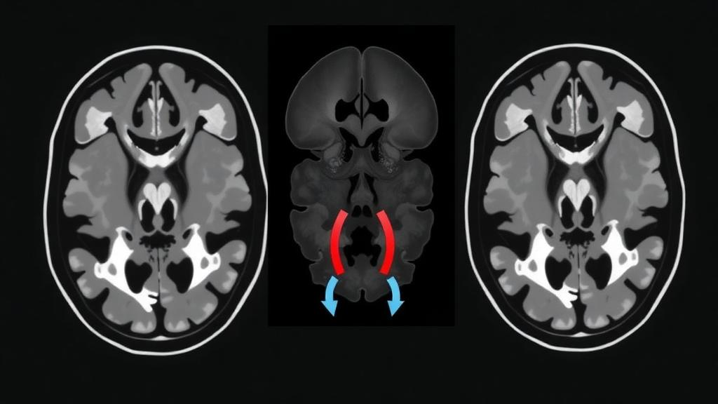
Researchers at Beijing Tiantan Hospital and the Beijing Neurosurgical Institute, affiliated with Capital Medical University, have developed and internally validated a set of machine-learning models that read quantitative texture patterns from routine CT angiography scans to distinguish symptomatic from asymptomatic unruptured spontaneous intracranial vertebrobasilar artery dissection, a condition abbreviated sIVBD. The retrospective single-center study, published in Frontiers in Neurology, included 173 patients and found that a combined model integrating clinical variables, conventional CTA features and a cross-fitted radiomics probability achieved the best overall performance among four prespecified models. Spontaneous intracranial vertebrobasilar artery dissection is characterized by disruption of the arterial wall with intramural hematoma formation, producing a spectrum of luminal and vascular wall abnormalities that includes stenosis, aneurysmal dilatation and other morphological changes. Its clinical presentation is highly variable, ranging from incidental or minimally symptomatic disease to posterior circulation ischemic stroke, subarachnoid hemorrhage and symptoms related to brainstem compression, which makes it an important and clinically challenging cerebrovascular disorder, particularly in young and middle-aged adults.
The natural history of sIVBD is similarly heterogeneous and remains difficult to predict. Some lesions may remain stable or improve spontaneously, whereas others may be associated with ischemic events due to hemodynamic compromise or thromboembolism, rupture with subarachnoid hemorrhage, or progressive enlargement with mass effect and neurological deterioration. This uncertainty has direct implications for management, because treatment decisions require integration of symptoms, lesion morphology, vascular anatomy and the potential risks of intervention, particularly since endovascular treatment of vertebrobasilar dissections may itself carry substantial procedural risk. Conventional clinical and imaging assessment remains essential but may not fully capture subtle lesion heterogeneity. Radiomics, the high-throughput extraction of quantitative imaging features, can capture lesion characteristics not readily appreciated by visual assessment and has increasingly been applied to intracranial vascular lesions, yet its utility for differentiating symptomatic from asymptomatic unruptured sIVBD on CT angiography had remained insufficiently investigated before this work.
The study cohort comprised 173 consecutive patients with unruptured sIVBD evaluated between May 2018 and September 2024, of whom 56 were symptomatic, presenting with ischemic neurological events, dissection-related pain or headache, or symptoms of local mass effect, and 117 were asymptomatic. Patients aged 18 to 75 years were eligible if they had a diagnosis established by CTA, digital subtraction angiography or MRI, pretreatment CTA images of sufficient quality, and adequate clinical information. Ruptured lesions, traumatic or iatrogenic dissections, coexisting intracranial tumors or vascular malformations, and prior treatment of the target lesion were excluded. The cohort was stratified by symptomatic status and randomly split at roughly 8:2 into a development cohort of 138 patients, with 93 asymptomatic and 45 symptomatic, and an independent internal validation cohort of 35 patients, with 24 asymptomatic and 11 symptomatic. In the overall cohort, symptomatic patients more often had hypertension, at 73.2 percent versus 49.6 percent, a history of cerebral infarction, at 37.5 percent versus 12.0 percent, higher modified Rankin Scale scores, basilar artery involvement, and larger lesions, with median lengths of 12.55 versus 8.36 millimeters and median widths of 7.06 versus 4.38 millimeters.
All CTA examinations were performed on a Revolution CT scanner using a standardized subtraction protocol, in which a noncontrast acquisition is deformably registered to the contrast-enhanced scan and subtracted, leaving a clean image of the opacified vessel lumen. Axial images were acquired at 80 kVp and 150 mA, reconstructed at 0.625 millimeter section thickness with an in-plane pixel spacing of approximately 0.49 by 0.49 millimeters, and contrast timing was individualized using a test-bolus technique with 15 milliliters of iodinated contrast injected at 6.0 milliliters per second before the 55-milliliter diagnostic acquisition. The resulting thin-section subtraction datasets were imported into 3D Slicer, where initial vascular segmentation was performed with a threshold-based approach followed by three-dimensional reconstruction and manual refinement. Three experienced neuroimaging specialists independently segmented every lesion, and the segmentation from the senior reader, with more than ten years of neurovascular imaging experience, was used for primary feature extraction, while the other two readers’ segmentations served to test reproducibility.
From each segmented lesion, the pipeline extracted 851 radiomic features using PyRadiomics: 14 shape descriptors plus first-order intensity statistics and texture features derived from gray-level co-occurrence, run-length, size-zone, dependence and neighboring gray-tone difference matrices, computed on both the original images and eight wavelet-decomposed versions. Of these, 722 features, or 84.8 percent, met the prespecified reproducibility criterion of an inter-reader intraclass correlation coefficient of at least 0.75, with a median ICC of 0.888. Feature selection then proceeded through Mann-Whitney U screening, Spearman correlation filtering at an absolute threshold of 0.90, minimum redundancy maximum relevance selection retaining up to 50 features, and finally LASSO logistic regression, which distilled the final full-development model to 20 features, predominantly wavelet-derived texture and first-order measures. Crucially, all outcome-informed selection was repeated independently within each training partition during validation, so no single feature set was allowed to leak across the evaluation boundary, although the outcome-independent ICC filter was determined once on the full development cohort before cross-validation began.
The investigators built four models. M1 used only conventional CTA morphology: lesion location, length, maximum width and parent artery stenosis. M2 added eight clinical variables, including age, sex, smoking, alcohol consumption, hypertension, diabetes, coronary artery disease and dyslipidemia. M3 was a radiomics-only model built on XGBoost, a gradient-boosted tree algorithm whose hyperparameters were tuned by a Grey Wolf Optimizer, a nature-inspired search method that evaluated 1,500 candidate configurations per outer partition, compared with 8,748 for grid search; the authors note that the two strategies used different search spaces and budgets, so the comparison does not establish superiority under equal computational resources. M4, the centerpiece, combined the twelve clinical and conventional CTA predictors with a single cross-fitted radiomics probability, meaning that each training patient’s radiomics score was generated by an upstream model that had never seen that patient’s outcome, a leakage-control strategy. All logistic models used ridge regularization, and a nomogram was produced to make the final combined model interpretable.
Primary evaluation used five-times-repeated five-fold stratified nested cross-validation, yielding 25 outer folds and one patient-averaged out-of-fold prediction per patient. In this rigorous development setting, the conventional CTA model achieved an area under the receiver operating characteristic curve of 0.630, the clinical-plus-CTA model 0.681, the radiomics-only model 0.676, and the combined model 0.723, which also posted the lowest Brier score at 0.193. In the prespecified primary comparison, adding radiomics to the clinical and conventional model increased the AUC by 0.0416, though the 95 percent confidence interval spanned zero and the paired DeLong p-value was 0.136, and it reduced the Brier score by 0.0101, with a bootstrap confidence interval of −0.0208 to 0.0006. Across the 25 outer folds, the combined model recorded both the highest mean AUC, 0.715, and the lowest between-fold variability, with a standard deviation of 0.0618, while the radiomics-only model showed the largest between-fold standard deviation at 0.0997.
The independent internal validation cohort of 35 patients, held out until every model and classification threshold had been locked, delivered striking numbers that demand careful reading. There, the combined model achieved an AUC of 0.966, with an average precision of 0.914 and a Brier score of 0.132, far outperforming the conventional models, which reached AUCs between 0.803 and 0.814. At the locked cutoff of 0.3167, the combined model correctly identified all 11 symptomatic patients, a sensitivity of 100 percent, while correctly classifying 19 of 24 asymptomatic patients, for a specificity of 79.2 percent and an accuracy of 85.7 percent. The positive predictive value was 68.8 percent and the negative predictive value was 100 percent. Yet the authors are explicit that this apparent leap in performance should not be taken at face value, because the validation cohort was small and its particular case mix may have contributed to the higher estimate.
The calibration analysis exposes that caution. Although the mean predicted probability in the validation cohort was close to the observed proportion of symptomatic patients, the calibration slope for the combined model was 6.4466, with a confidence interval from 2.9194 to 13.4592, indicating that the predicted log odds were insufficiently dispersed and that individual probability estimates were not reliable. An AUC measures only how well a model ranks patients, and it can remain high even when the actual probabilities are badly miscalibrated. The authors therefore state that the predicted probabilities should not be used directly for individual clinical decision-making without evaluation in a larger external cohort and, if necessary, recalibration. The gap between development and validation AUCs, together with the imprecisely estimated calibration parameters, may reflect sampling variability in a validation cohort containing only 35 patients and 11 symptomatic events.
Interpretability and stability analyses added further texture to the story. SHAP analysis of the final radiomics model identified wavelet-HHH.glszm.ZoneVariance, a measure of gray-level variability within uniformly sized zones at the finest wavelet scale, as the leading contributor, with a mean absolute SHAP value of 0.4481, followed by wavelet-LLL.gldm.LargeDependenceLowGrayLevelEmphasis and wavelet-LHH.glszm.SizeZoneNonUniformityNormalized. These features quantify spatial heterogeneity in the vessel wall image beyond simple geometry, and notably the shape descriptor Maximum2DDiameterSlice showed minimal correlation with manually recorded lesion length and width, suggesting radiomics offers a mathematically distinct representation of lesion form. However, SHAP values describe how the fitted model uses features, not evidence that they are causal determinants of symptomatic presentation. Feature selection itself was unstable: no individual feature appeared in all 25 training partitions, the most frequently retained feature appeared in 17, and the mean pairwise Jaccard similarity between selected sets was only 0.1493, a pattern the authors attribute to many correlated features encoding overlapping information. The limitations are candidly acknowledged: the study was retrospective and single-center, the development cohort contained only 45 symptomatic patients, manual segmentation remains a potential source of measurement variability, treatment allocation was not randomized, and the cross-sectional design means the models characterize symptomatic status at the time of assessment rather than predicting future enlargement, rupture or treatment response. Larger multicenter studies with true external validation will determine whether CTA radiomics can complement, rather than replace, conventional assessment of this uncertain cerebrovascular disease.
Subject of Research: Development and internal validation of CTA-based radiomics models for differentiating symptomatic from asymptomatic unruptured spontaneous intracranial vertebrobasilar artery dissection
Article Title: Development and internal validation of CTA-based radiomics models for differentiating symptomatic from asymptomatic unruptured spontaneous intracranial vertebrobasilar artery dissection
Article References: Jiang, J., Bi, S., Gui, S., Zheng, L., Li, Y., & Liu, Q. (2026). Development and internal validation of CTA-based radiomics models for differentiating symptomatic from asymptomatic unruptured spontaneous intracranial vertebrobasilar artery dissection. Frontiers in Neurology, 17, Article 1883223. https://doi.org/10.3389/fneur.2026.1883223
Image Credits: AI Generated
DOI: 10.3389/fneur.2026.1883223
Keywords: radiomics, CT angiography, vertebrobasilar artery dissection, machine learning, XGBoost, prediction models, neurology, stroke, Development, internal, validation, CTA-based
Cite Scienmag News
Ophelia Keating. (October 24, 2026). CTA Radiomics Model Distinguishes Symptomatic From Silent Vertebrobasilar Dissections. Scienmag. https://scienmag.com/cta-radiomics-model-distinguishes-symptomatic-from-silent-vertebrobasilar-dissections/
Ophelia Keating. "CTA Radiomics Model Distinguishes Symptomatic From Silent Vertebrobasilar Dissections." Scienmag, 24 October 2026, https://scienmag.com/cta-radiomics-model-distinguishes-symptomatic-from-silent-vertebrobasilar-dissections/. Accessed 24 October 2026.
Ophelia Keating. "CTA Radiomics Model Distinguishes Symptomatic From Silent Vertebrobasilar Dissections." Scienmag. October 24, 2026. https://scienmag.com/cta-radiomics-model-distinguishes-symptomatic-from-silent-vertebrobasilar-dissections/