


A short correspondence published in the Journal of Neuro-Oncology is making a pointed argument that could reshape how neuro-oncologists interpret a familiar group of brain tumors. Writing in the journal’s September 2026 issue, neurosurgeons Alexandre Roux, Marc Zanello and Johan Pallud of GHU-Paris Psychiatrie et Neurosciences, Hôpital Sainte Anne, contend that contrast enhancement on magnetic resonance imaging is the missing imaging variable in IDH-wildtype gliomas that carry a historical World Health Organization grade 2 or grade 3 histology. Their argument arrives as a direct commentary on a companion study in the same journal by Carriere and colleagues, which reported long-term outcomes for this diagnostically awkward population of patients.
To understand why the correspondence matters, it helps to unpack the diagnostic category at its center. The 2021 fifth edition of the WHO classification of tumors of the central nervous system reorganized diffuse gliomas around molecular features, most importantly mutations in the isocitrate dehydrogenase genes. Under the new framework, a diffuse astrocytic tumor without an IDH mutation is classified as astrocytoma, IDH-wildtype, and in adults this molecular profile places the tumor in the same family as glioblastoma. That shift left a residue of tumors that were diagnosed years ago as grade 2 or grade 3 diffuse astrocytomas on histology alone, before molecular testing was routine, but which are now understood to be IDH-wildtype. Carriere and colleagues examined the long-term outcomes of exactly this group, and their findings carry weight for the many patients whose archived diagnoses are being retrospectively reinterpreted.
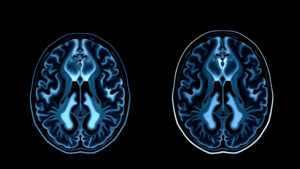
The Paris authors’ central claim is that the companion study, and much of the literature surrounding it, has overlooked a variable that has been sitting in plain sight on every diagnostic scan: whether the tumor takes up gadolinium contrast. On a contrast-enhanced MRI, gadolinium normally stays within blood vessels because the blood-brain barrier keeps large molecules out of brain tissue. When a tumor disrupts that barrier, contrast leaks into the lesion and the region lights up on T1-weighted images. That enhancement is not a random artifact; it is a macroscopic signature of vascular proliferation and barrier breakdown, processes that correspond to aggressive biological behavior. In glioma practice, enhancement has long been one of the most scrutinized features on any scan.
The evidence base for that scrutiny is substantial, and the correspondence anchors its argument in three prior studies from overlapping author groups. The earliest, published by Pallud and colleagues in Neuro-Oncology in 2009, examined the prognostic significance of imaging contrast enhancement in WHO grade II gliomas and found that enhancement carried prognostic weight in these low-grade tumors. A second study, published by Roux and colleagues in Brain Pathology in 2020, asked whether adding MRI data improved the prognostic models built on the WHO 2016 classification and the cIMPACT-NOW updates for diffuse astrocytic tumors in adults, working toward the extended use of MRI data in integrated glioma diagnosis. A third, published by Roux and colleagues in the Journal of Neuro-Oncology in 2024, demonstrated the prognostic significance of MRI contrast enhancement in newly diagnosed glioblastoma, IDH-wildtype, according to the WHO 2021 classification.
Read together, those three studies trace a consistent thread across the modern diagnostic eras. Enhancement matters in tumors that look low-grade under the microscope. It matters when imaging features are layered on top of molecular and histological classifications to refine prognosis. And it matters even within glioblastoma, IDH-wildtype, where all patients already carry the worst prognostic label in neuro-oncology and yet enhancement still separates groups with different outcomes. The correspondence argues that the logical next step is to apply the same lens to the historical grade 2 and grade 3 IDH-wildtype gliomas that Carriere and colleagues analyzed, because these tumors sit precisely at the intersection where the prior evidence converges.
The stakes of that omission are easiest to see in the numbers that clinicians quote at the bedside. Historical grade 2 and grade 3 diffuse astrocytomas were traditionally described as indolent or moderately aggressive tumors with survival measured in many years, and counseling for newly diagnosed patients was built on those expectations. IDH-wildtype biology erodes that optimism considerably, because tumors in this molecular group behave more like glioblastomas even when their histology appears lower grade. If a patient’s tumor is reclassified retrospectively as IDH-wildtype astrocytoma, the prognostic conversation changes dramatically. But within that reclassified population, outcomes are unlikely to be uniform, and the correspondence suggests that contrast enhancement is one of the variables most likely to capture that heterogeneity.
The biological rationale for expecting enhancement to stratify these tumors is straightforward. Enhancement reflects disruption of the blood-brain barrier and, in gliomas, is closely associated with microvascular proliferation, one of the histological hallmarks that traditionally pushed a tumor toward a higher grade. A histologically grade 2 or grade 3 appearing tumor that nonetheless shows contrast enhancement is, in effect, displaying imaging evidence of aggressive vascular biology that the microscope may have undersampled. Diffuse gliomas are notoriously heterogeneous, and a small surgical specimen can miss foci of higher-grade biology elsewhere in the lesion. Enhancement on MRI offers a whole-tumor view that histology from a biopsy or even a resection cannot match, which is precisely why the authors describe it as a missing variable rather than a redundant one.
There is also a practical dimension to the argument that extends beyond prognosis. Treatment decisions for diffuse lower-grade gliomas, including the timing of surgery, the extent of resection attempted, and the threshold for adding radiotherapy and chemotherapy, are calibrated to expected tumor behavior. If enhancement identifies a subset of histologically lower-grade, IDH-wildtype tumors that behave like glioblastomas, those patients might warrant more intensive surveillance and earlier aggressive treatment, while patients with non-enhancing tumors might safely remain on the watch-and-wait pathways that have transformed quality of life in genuinely indolent glioma care. Conversely, trials that enroll historical grade 2 and 3 IDH-wildtype tumors as a single cohort risk diluting their results if enhancement status is not recorded and controlled for, because the cohort would mix biologically distinct populations.
The correspondence also lands at a moment when the field is actively debating how much imaging should be formalized in glioma classification. The 2021 WHO edition made molecular markers the backbone of diagnosis, but it stopped short of requiring specific imaging features, leaving radiological findings as contextual information rather than diagnostic criteria. Studies such as the 2020 Brain Pathology work argue that MRI data add prognostic value on top of molecular and histological categories, and the 2024 glioblastoma study shows that even within a single molecular class, imaging separates meaningful subgroups. The Paris authors’ commentary pushes that logic one step further: for the growing population of retrospectively reclassified IDH-wildtype tumors, imaging variables may be the most practical tool available to recover prognostic granularity that histology alone no longer provides.
None of this diminishes the contribution of the Carriere study, which the correspondence treats as a valuable foundation rather than a flawed analysis. Long-term outcome data for historical grade 2 and 3 IDH-wildtype gliomas are exactly what the field needs as archived diagnoses are revisited under the molecular era’s framework. The authors’ point is additive: those outcomes should be reanalyzed, and future cohorts should be analyzed from the start, with contrast enhancement as an explicit stratification variable. As hospitals increasingly re-review historical specimens with modern molecular panels, thousands of patients will find themselves in this reclassified category, and the questions their clinicians ask will hinge on which prognostic variables were captured. A variable as routinely available as contrast enhancement, the correspondence argues, should not be the one that gets away.
Subject of Research: Prognostic value of MRI contrast enhancement in IDH-wildtype gliomas with historical WHO grade 2 and 3 histology
Article Title: Contrast enhancement: the missing imaging variable in IDH-wildtype gliomas with historical WHO grade 2 and 3 histology
Article References: Roux, A., Zanello, M., & Pallud, J. (2026). Contrast enhancement: the missing imaging variable in IDH-wildtype gliomas with historical WHO grade 2 and 3 histology. Journal of Neuro-Oncology, 179(2), Article 74. https://doi.org/10.1007/s11060-026-05791-1
Image Credits: AI Generated
DOI: 10.1007/s11060-026-05791-1
Keywords: glioma, IDH-wildtype, contrast enhancement, MRI, WHO classification, WHO grade 2, WHO grade 3, prognosis, neuro-oncology, blood-brain barrier, glioblastoma, neuroradiology
Cite Scienmag News
Nathaniel Bowman. (October 2, 2026). Contrast Enhancement on MRI May Be the Missing Prognostic Variable in IDH-Wildtype Gliomas Diagnosed as Grade 2 and 3. Scienmag. https://scienmag.com/contrast-enhancement-on-mri-may-be-the-missing-prognostic-variable-in-idh-wildtype-gliomas-diagnosed-as-grade-2-and-3/
Nathaniel Bowman. "Contrast Enhancement on MRI May Be the Missing Prognostic Variable in IDH-Wildtype Gliomas Diagnosed as Grade 2 and 3." Scienmag, 2 October 2026, https://scienmag.com/contrast-enhancement-on-mri-may-be-the-missing-prognostic-variable-in-idh-wildtype-gliomas-diagnosed-as-grade-2-and-3/. Accessed 4 October 2026.
Nathaniel Bowman. "Contrast Enhancement on MRI May Be the Missing Prognostic Variable in IDH-Wildtype Gliomas Diagnosed as Grade 2 and 3." Scienmag. October 2, 2026. https://scienmag.com/contrast-enhancement-on-mri-may-be-the-missing-prognostic-variable-in-idh-wildtype-gliomas-diagnosed-as-grade-2-and-3/


