Forensic documentation of facial and neck injuries may be on the verge of a practical upgrade. In a technical feasibility study published in the Egyptian Journal of Forensic Sciences, researchers report that a compact, single-capture 3D imaging system originally built for aesthetic medicine and dermatology successfully generated photorealistic, metrically scaled 3D surface models of the face and neck in simulated forensic scenarios. The entire image acquisition took less than one second, and fully automated reconstruction was available within roughly 30 seconds on a laptop computer or up to two minutes on a tablet device, depending on the processing hardware used.
Three adult volunteers underwent 3D facial scanning after simulated injury patterns were applied to their skin. These included temporary trauma tattoo markers simulating abrasions, contusions and patterned blunt force injuries, as well as dermatological red marker drawings simulating petechial hemorrhages of the kind relevant in suspected strangulation cases. The assessment was deliberately descriptive and qualitative rather than a formal accuracy validation.
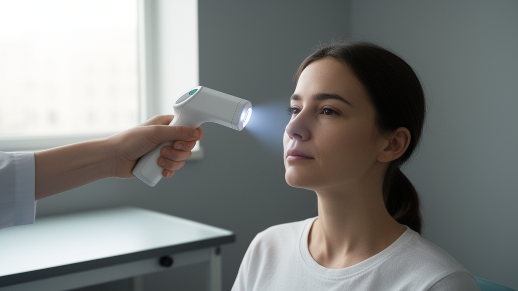
The hardware behind the system is a synchronized multi-camera array of 13 CMOS sensor cameras, each with a resolution of up to 13 megapixels, combined with 18 white LED light units operating at a color temperature of 4000 kelvin. The illumination incorporates polarization-controlled lighting, which enables both specular and diffuse reflection imaging. All cameras are triggered simultaneously, so the facial surface is captured in a single shot that minimizes motion artifacts. According to the manufacturer specifications reported in the study, the root mean square accuracy of the measurement points is 0.1 millimeters. Proprietary software then automatically performs photogrammetric reconstruction, mesh generation and texture mapping, producing a textured 3D surface model of the face and neck without any external software or manual post-processing.
In all three subjects, the system produced a complete 3D surface model of the face and cervical region from a single capture. Reconstruction was achieved without visible stitching artifacts, surface discontinuities or misalignment, even in anatomically complex regions such as the periorbital area, nasal contours, auricular margins and the mandibular border. The resulting models combined high-resolution surface geometry with photorealistic texture mapping, preserving skin tone, pigmentation patterns and fine surface structures including wrinkles and pores. One of the volunteers was an older adult with more pronounced baseline wrinkles and skin texture variations, and injury visualization remained consistent across all subjects, with age-related skin features not interfering with the identification or delineation of the simulated trauma patterns.
The simulated injuries themselves were clearly represented on the reconstructed models. Temporary tattoo-based abrasions, contusions and patterned injuries were visualized with preserved shape, border characteristics, color distribution and anatomical localization. The marker-based petechial simulations in the cervical region were detectable both in standard texture rendering and in vascularity-sensitive visualization layers. Beyond the standard photorealistic rendering, the system offers texture-based, pigmentation-sensitive and vascularity-sensitive visualization modes. Texture-based layers enhanced the depiction of surface irregularities and erythematous regions, pigmentation-sensitive layers highlighted contrast variations, and vascularity-sensitive layers increased the visibility of the simulated petechial patterns, providing complementary perspectives to conventional photography-like rendering.
Measurement functionality also performed as intended in this feasibility setting. Linear distance measurements and angular assessments could be performed directly on the 3D models, and the system allowed assessment of spatial relationships between injury patterns and anatomical reference points, including bilateral comparisons. No measurement instability or clinically relevant mesh deformation was observed during interactive manipulation of the injury regions assessed. A laterally positioned rectangular reference scale in the study images confirmed concordance between virtual measurements and corresponding physical dimensions. The authors note, however, that minor localized reconstruction artifacts may occur in anatomically challenging regions such as the ocular surface, a caveat that matters when interpreting delicate morphometric landmarks.
Practical handling was another focus of the evaluation. Subjects were positioned upright in a neutral head position with relaxed facial musculature, and one single-capture acquisition was performed per subject with no repeated scans or manual post-acquisition adjustments. To simulate realistic forensic conditions, no surface preparation such as make-up correction or skin treatment was applied. No motion artifacts were observed, reconstruction was fully automated and required no manual correction, and all subjects tolerated the procedure without discomfort. The qualitative assessment was carried out by a board-certified forensic pathologist with more than 20 years of experience in forensic 3D surface documentation, photogrammetry and three-dimensional visualization.
The context for this work is a long-standing gap in forensic practice. Although 3D surface documentation offers clear conceptual advantages, routine documentation of external injuries still relies predominantly on two-dimensional photography, which flattens complex anatomical structures into a planar representation and can obscure depth relationships, geometric configuration and the spatial alignment of patterned injuries. This may limit objective tool-wound comparison and complicate expert review in cases of blunt force trauma, patterned injuries or suspected strangulation. Early forensic optical surface digitizing demonstrated that photogrammetry and structured-light scanning can generate color-true, metrically scaled 3D models, and these approaches later evolved into multimodal workflows integrating surface scans with computed tomography and magnetic resonance imaging, forming the basis of comprehensive digital forensic reconstruction concepts such as Virtopsy. But many established systems require multi-step acquisition protocols, complex calibration or stationary installations, restricting their use in time-sensitive or space-limited environments such as emergency departments, forensic outpatient clinics, police facilities or pre-autopsy documentation settings.
The mugshot angle adds a further dimension. The classical police identification photograph has consisted of standardized frontal and profile views since the nineteenth century, and in recent years this procedure has increasingly been complemented by 3D acquisition techniques aimed at generating complete facial models that can be freely rotated and viewed from varying angles. Seminal work by Leipner and colleagues described a photogrammetric multi-camera system for generating forensic 3D head models, and a 2024 study by Giuliani and colleagues examined a multi-vision system designed to enhance forensic facial identification. Both showed that 3D models offer advantages over conventional two-dimensional mugshots, particularly when comparison images are captured from non-frontal perspectives. The authors of the present study suggest that 3D mugshots should be regarded not as a replacement but as a technological extension of traditional police photographic documentation, and they note potential applications including automated comparison with surveillance footage, reconstruction of facial views from limited image sources, retrospective derivation of frontal, lateral and oblique projections from a single metrically scaled model, and integration with biometric databases. Any implementation in police identification workflows, they caution, would require independent validation, regulatory assessment and evaluation of biometric interoperability, and data protection issues around the storage of biometric information are increasingly central to scientific and legal discourse.
The limitations of the study are substantial and the authors state them plainly. Only three volunteers were included, all injuries were simulated, and the simulated patterns did not include substantial depth-related surface alterations such as depressions, lacerations or pronounced patterned compression marks. No quantitative geometric accuracy validation against external reference standards was performed, since the primary aim was technical feasibility rather than metric validation. Consequently, no conclusions can be drawn about diagnostic sensitivity, specificity or comparative superiority relative to established forensic imaging systems. The authors also discuss, but did not test, the possibility of adapting the compact device to horizontal examination settings, such as positioning it above or laterally adjacent to a supine body for pre-autopsy 3D documentation of facial and cervical trauma before soft tissue dissection; this application was not experimentally evaluated under postmortem conditions, and practical handling, image quality and reproducibility in mortuary settings remain to be systematically investigated. They position compact single-capture systems as complementary to larger stationary full-body installations rather than competing with them, noting that a recent feasibility study of the dermatology-based VECTRA WB360 system demonstrated that full-body scanning combined with AI-supported lesion detection can enhance standardization in forensic documentation. Formal validation under clinical and postmortem conditions, the team concludes, remains necessary before any routine implementation of compact single-shot facial 3D imaging in forensic workflows.
Subject of Research: Technical feasibility of adapting a compact single-capture 3D facial imaging system for forensic documentation of simulated face and neck injuries
Article Title: Single-capture 3D mugshot imaging of the face and neck: adapting a compact mobile system for forensic documentation
Article References: Thali, M. J., Thali, M. H., & Gascho, D. (2026). Single-capture 3D mugshot imaging of the face and neck: adapting a compact mobile system for forensic documentation. Egyptian Journal of Forensic Sciences, 16(1), Article 79. https://doi.org/10.1186/s41935-026-00578-x
Image Credits: AI Generated
DOI: 10.1186/s41935-026-00578-x
Keywords: forensic imaging, 3D surface scanning, photogrammetry, facial injury documentation, mugshot, single-capture acquisition, trauma documentation, feasibility study, Virtopsy, biometrics, forensic medicine, 3D reconstruction
Cite Scienmag News
Ophelia Keating. (October 30, 2026). Compact 3D Scanner Captures Face and Neck Injuries in Under One Second. Scienmag. https://scienmag.com/compact-3d-scanner-captures-face-and-neck-injuries-in-under-one-second/
Ophelia Keating. "Compact 3D Scanner Captures Face and Neck Injuries in Under One Second." Scienmag, 30 October 2026, https://scienmag.com/compact-3d-scanner-captures-face-and-neck-injuries-in-under-one-second/. Accessed 31 October 2026.
Ophelia Keating. "Compact 3D Scanner Captures Face and Neck Injuries in Under One Second." Scienmag. October 30, 2026. https://scienmag.com/compact-3d-scanner-captures-face-and-neck-injuries-in-under-one-second/