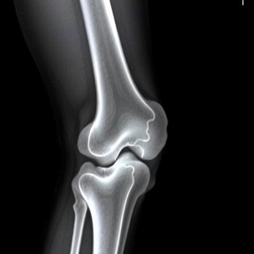
Recent advancements in radiological research have focused on the intricate variations that occur in the metaphyseal regions of long bones, particularly within the distal femurs and proximal tibias. This observational study illuminates the vital importance of accurately distinguishing these subtle anatomical differences from classic metaphyseal lesions often associated with pediatric pathologies. The research, conducted by a team led by Karmazyn et al., lays the groundwork for further exploration in diagnosing conditions involving these critical areas of the skeletal system.
Metaphyseal lesions are typically associated with conditions such as metabolic bone diseases, trauma, and various malignancies. Understanding their anatomical nuances is crucial for clinicians, as misinterpretation can lead to misdiagnosis and inappropriate management strategies. The study makes a compelling argument for the need to refine diagnostic criteria that separate the variations specific to normal anatomy from those that indicate pathology. This improved understanding will significantly impact clinical practices, particularly in pediatric radiology.
One noteworthy aspect of the study is its radiological methodologies, which utilize advanced imaging techniques capable of providing detailed insight into bone morphology. These methods enable researchers to identify and classify the features of metaphyseal regions more precisely. High-resolution imaging, alongside quantitative analysis techniques, enhances the reliability of the findings reported in the study. It emphasizes the necessity for radiologists to engage with evolving technology in their diagnostic workflow.
The study draws on a sizeable cohort of pediatric patients, making it suitable for a robust statistical analysis. The diversity in demographic factors such as age, sex, and underlying health conditions provides a comprehensive overview of how metaphyseal variations manifest in different populations. This extensive dataset fuels the argument that the recognition of normal anatomical variations is as essential as identifying lesions, allowing for a more precise interpretation of pediatric radiology images.
As bone development varies significantly among children as they grow, the study underscores the need for age-appropriate references for metaphyseal architecture. This vital information aids practitioners in formulating age-specific diagnostic approaches that acknowledge the inherent biological diversity within the pediatric population. Armed with this knowledge, healthcare providers can enhance their clinical acumen when interpreting imaging studies, especially in cases where there exists a clinical suspicion of pathologic processes.
Furthermore, the implications extend to educational paradigms within medicine and radiology. Training programs might incorporate these findings to foster a nuanced understanding among future practitioners. By highlighting the importance of distinguishing normal from pathological findings, the research contributes to shaping curricula that address the complexities of interpreting pediatric imaging studies.
The potential for a paradigm shift toward better diagnostic clarity is promising. Enhanced recognition of metaphyseal variations might contribute to diminishing the rate of unnecessary interventions tied to misdiagnosed conditions. The research aligns with a broader trend in medicine that advocates for precision and personalization in healthcare, ensuring that the variations inherent to normal human anatomy are respected and understood.
Critically, this study opens up avenues for further research. Future investigations could expand on these findings, exploring how these metaphyseal characteristics change with age and health status. Longitudinal studies might solidify these findings, establishing a framework for understanding the natural history of these anatomical variations.
The research also raises questions regarding the role of genetic and environmental factors in shaping metaphyseal morphology. These dimensions are vital to explore as they could potentially unveil underlying mechanisms driving both normal and pathological skeletal development. By bridging genetic research with clinical radiology, a more holistic understanding of bone disorders may emerge.
In conclusion, Karmazyn et al.’s study serves as a groundbreaking exploration into the nuanced world of metaphyseal anatomy within pediatric patients. By distinguishing between normal variations and lesions, this research is poised to enhance diagnostic accuracy, support better clinical decisions, and ultimately improve patient outcomes. As medical imaging continues to advance, the findings from this study will likely resonate throughout the fields of pediatrics and radiology, paving the way for a future where the interplay between intricate anatomical knowledge and clinical practice leads to more effective healthcare.
In this rapidly evolving medical landscape, maintaining an awareness of such advancements is critical. Clinicians who remain informed will be better equipped to interpret their findings accurately and apply them to patient care. As evidenced by the compelling case made by Karmazyn et al., the integration of radiological insights with clinical practice can transform how we approach pediatric bone diseases. With this foundational knowledge in hand, the medical community is empowered to refine its approach to diagnosing and treating conditions related to the skeletal system.
As the study continues to receive attention in academic circles, the relevance of its findings is likely to expand beyond the initial scope. Medical professionals, educators, and researchers alike will benefit from incorporating these insights into their work, ultimately fostering a more sophisticated understanding of pediatric radiology. Hence, it is critical to adopt these innovations and insights for the ongoing evolution of medical imaging and diagnosis.
Strong advocacy for continued education in this domain, interprofessional collaboration, and evolving technology will ultimately lead to enhanced patient care. The refinement of diagnosis not only caters to the immediate needs of the patient but also lays the groundwork for future research consonant with advancing our comprehension of bone health in pediatrics.
As we delve deeper into the significance of this research, it becomes apparent that the excitement rests in how individual variations are now seen as a critical component rather than mere outliers. By embracing the complexities of metaphyseal variations, the field of pediatrics and radiology can progress towards a more accurate and personalized standard of care, demonstrating the necessity of ongoing research to inform clinical practice effectively.
Subject of Research: Metaphyseal variations in distal femurs and proximal tibias
Article Title: Can metaphyseal variations in the distal femurs and proximal tibias be distinguished from classic metaphyseal lesions?
Article References:
Karmazyn, B., Newman, C.L., Marine, M.B. et al. Can metaphyseal variations in the distal femurs and proximal tibias be distinguished from classic metaphyseal lesions? Pediatr Radiol (2025). https://doi.org/10.1007/s00247-025-06398-w
Image Credits: AI Generated
DOI: https://doi.org/10.1007/s00247-025-06398-w
Keywords: Metaphyseal lesions, pediatric radiology, bone morphology, diagnostic accuracy, skeletal development.


