


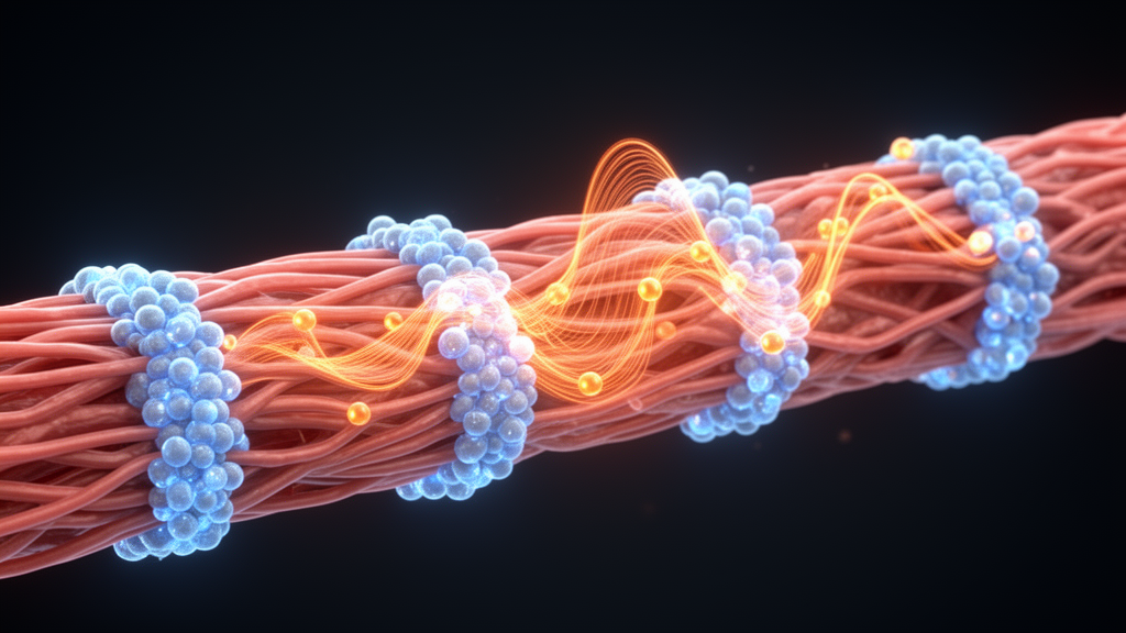
Calcium waves that sweep across heart muscle cells depend on a surprisingly specific molecular arrangement, according to a new computational study. Using a three-dimensional model of mouse ventricular myocytes built at submicrometer resolution, researchers found that the calcium-binding protein calsequestrin2 (CASQ2) must sit alongside ryanodine receptor type 2 (RyR2) clusters for waves to travel reliably through the cell.
Cardiac calcium waves propagate through a fire-diffuse-fire mechanism: calcium released from one RyR2 cluster diffuses to neighboring clusters and triggers their activation. This process faces a fundamental physical challenge. The signal must cross distances of 1 to 2 micrometers between Z-planes while being weakened by cytosolic buffering and diffusion, and at the same time each release site depletes its local sarcoplasmic reticulum calcium store. How waves manage to propagate efficiently despite these constraints has remained unclear.
The modeling revealed a clear answer. In a physiological configuration, where CASQ2 is concentrated at release sites as observed experimentally, calcium waves propagated reliably across the cell, with velocities matching the experimental range. When the researchers redistributed the same total amount of CASQ2 uniformly throughout the sarcoplasmic reticulum, the wavefront stalled. Keeping the protein’s overall quantity unchanged but altering only its localization was enough to abolish propagation.
These results identify CASQ2–RyR2 co-localization as a key structural requirement for effective calcium wave propagation in ventricular myocytes. Because the study is based on a computational model of mouse ventricular cells rather than direct experimental measurements in human tissue, its conclusions describe model predictions grounded in experimentally observed protein distributions. Within that scope, the findings indicate that the spatial organization of calcium stores at release sites, not merely the total amount of buffering protein, determines whether disturbances in cellular calcium can spread as coordinated waves.
Subject of Research: Calsequestrin localization at RyR2 clusters enables calcium wave propagation in ventricular myocytes
Article Title: Calsequestrin localization at RyR2 clusters enables calcium wave propagation in ventricular myocytes
Article References: Conesa, D., Echebarria, B., Hove-Madsen, L., Shiferaw, Y., & Alvarez-Lacalle, E. (2026). Calsequestrin localization at RyR2 clusters enables calcium wave propagation in ventricular myocytes. PLOS Computational Biology, 22(10), e1014449. https://doi.org/10.1371/journal.pcbi.1014449
Image Credits: AI Generated
DOI: 10.1371/journal.pcbi.1014449
Keywords: Calsequestrin, localization, RyR2, clusters, enables, calcium, wave, propagation, ventricular, myocytes, scientific research
Cite Scienmag News
Drew Townsend. (October 26, 2026). Calsequestrin clustering powers calcium waves in heart cells. Scienmag. https://scienmag.com/calsequestrin-clustering-powers-calcium-waves-in-heart-cells/
Drew Townsend. "Calsequestrin clustering powers calcium waves in heart cells." Scienmag, 26 October 2026, https://scienmag.com/calsequestrin-clustering-powers-calcium-waves-in-heart-cells/. Accessed 26 October 2026.
Drew Townsend. "Calsequestrin clustering powers calcium waves in heart cells." Scienmag. October 26, 2026. https://scienmag.com/calsequestrin-clustering-powers-calcium-waves-in-heart-cells/


