


Spinal cord injury is one of medicine’s most stubborn problems. Nearly every patient with a complete injury, and most with incomplete injuries, live with lasting motor deficits, and roughly two-thirds suffer chronic neuropathic pain, a burning, electric torment that can make rehabilitation almost impossible. Complete neurological recovery is exceptionally rare, occurring in fewer than one percent of cases. Now a translational study published in Experimental & Molecular Medicine offers something the field has lacked for decades: a precise, cell-by-cell explanation of how a noninvasive brain stimulation technique relieves both the paralysis and the pain at once, and a single molecular target that appears to be indispensable for the therapy to work.
The technique in question is high-frequency repetitive transcranial magnetic stimulation, or HF-rTMS, in which rapidly pulsed magnetic fields are applied over the primary motor cortex, the brain region that commands voluntary movement. Clinicians have long observed that this stimulation can improve motor recovery and dull neuropathic pain, but the two effects were almost always studied separately, and the cellular mechanism remained a black box. A team led by researchers at Xijing Hospital and the Fourth Military Medical University in Xi’an, China, set out to open that box, combining retrospective clinical data, a mouse model of spinal cord injury, single-nucleus RNA sequencing, electron microscopy, and viral gene therapy into one unusually complete causal chain.
The clinical evidence came first. The team reviewed records of 93 patients with spinal cord injury admitted between January 2018 and June 2025, of whom 40 received four consecutive weeks of HF-rTMS targeted at the left primary motor cortex at 10 Hz, 1,200 pulses per session. Compared with patients who did not receive stimulation, the treated group showed significantly greater gains on the modified Barthel index and manual muscle test, and, strikingly, only the stimulated group achieved a significant reduction in pain on the numerical rating scale. One therapy, two symptom domains, improved simultaneously in the same patients.
To understand why, the researchers turned to mice. They inflicted a standardized compression injury at the tenth thoracic vertebra, which produced lasting hind limb motor impairment measured by the Basso Mouse Scale, alongside mechanical allodynia and thermal hyperalgesia measured with von Frey filaments and a thermal testing apparatus. High-speed gait analysis on a glass floor added objective detail: injured mice walked more slowly, with worse coordination, and visibly shifted their weight away from painful hind paws. Five weeks of daily HF-rTMS, starting three days after injury, accelerated motor recovery, reversed both measures of pain hypersensitivity, and restored normal weight-bearing in the gait analysis. The mouse model faithfully reproduced the human motor-pain comorbidity, and the stimulation relieved both.
The mechanistic hunt then went molecular. The team sequenced roughly 90,000 individual nuclei from the primary motor cortex of uninjured, injured, and injured-and-stimulated mice, sorting them into nine major cell types. When they asked which biological processes were disrupted by injury and then reversed by stimulation, one theme dominated: mitochondrial energy metabolism, including oxidative phosphorylation, ATP synthesis coupled to electron transport, and aerobic respiration. And the reversal was strongest not in the excitatory neurons that send motor commands down the spinal cord, but in GABAergic inhibitory interneurons, the cells that keep cortical circuits balanced and prevent runaway excitation.
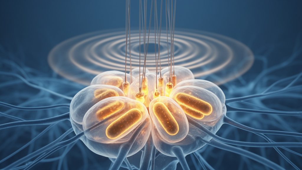
Mitochondria are the cell’s power plants, and their health depends on a constant cycle of fission and fusion. The master regulator of fission is a protein called dynamin-related protein 1, or Drp1, which pinches damaged mitochondria into smaller pieces so they can be repaired or cleared. Using transmission electron microscopy, the researchers identified GABAergic synapses in layer V of the motor cortex by their symmetric structure and pleomorphic vesicles, then measured the mitochondria inside those neurons. After injury, the mitochondria grew progressively larger and sparser, and their circularity, a marker linked to mitochondrial toxicity, climbed over five weeks. Western blots confirmed a rapid, sustained drop in Drp1 protein in the motor cortex, while the fusion proteins mitofusin 1 and 2 were untouched. The result was a bioenergetic crisis: ATP levels in the cortex fell sharply, and markers of damaged mitochondrial accumulation, VDAC-1 and Tom20, rose.
Here the story becomes remarkable in its specificity. Confocal imaging showed that the Drp1 loss occurred selectively within GAD67-positive GABAergic neurons, not in neighboring glutamatergic neurons, and that five weeks of HF-rTMS restored Drp1 expression only in that same inhibitory population. Electron microscopy confirmed that stimulation normalized mitochondrial size, density, and circularity in GABAergic neurons while leaving glutamatergic mitochondria largely unchanged. Cell-type-specific fluorescent staining for mitochondrial superoxide and the ATP synthase subunit ATP5A revealed elevated oxidative stress and diminished energy production confined to the inhibitory neurons, defects that both stimulation and Drp1 restoration reversed. Notably, the primary somatosensory cortex, a region deeply involved in pain perception, showed no such Drp1 response to stimulation, which may explain why clinical rTMS protocols aimed at the motor cortex tend to outperform those aimed at the sensory cortex for analgesia.
Correlation, however, is not causation, and this is where the study sets a new bar for neuromodulation research. Using adeno-associated viruses driven by the GAD67 promoter, the team overexpressed Drp1 exclusively in GABAergic neurons of the motor cortex. In injured mice, this single genetic intervention mimicked the entire therapeutic package: better motor scores from week two onward, reduced allodynia and hyperalgesia, improved gait, normalized mitochondrial ultrastructure, and ATP levels pushed back toward normal. Conversely, knocking Drp1 down in those same neurons worsened the injury phenotype and, most decisively, completely abolished every benefit of HF-rTMS. A pharmacological inhibitor of Drp1, Mdivi-1, likewise nullified the stimulation’s effects on motor function and pain. Drp1 in layer V GABAergic neurons of the motor cortex is thus both necessary and sufficient for the therapy to work, a bidirectional mediator of rTMS responsiveness.
Why would inhibitory interneurons be the vulnerable population? GABAergic neurons make up only about 15 to 20 percent of cortical cells but carry enormous metabolic demands, and spinal cord injury is known to reduce GABAergic inhibition in sensory and motor cortices, a disinhibition that contributes to both maladaptive motor plasticity and chronic pain. If Drp1 deficiency starves these neurons of ATP, their energy-intensive firing falters, cortical excitatory-inhibitory balance tips, and the motor-pain comorbidity follows. By restoring fission and bioenergetics in this microcircuit, HF-rTMS may re-arm the inhibitory system and, through the motor cortex’s descending connections to the periaqueductal gray and zona incerta, re-engage the brain’s endogenous pain-control machinery. The finding also carries a practical warning: common drugs that impair mitochondrial fission, including olanzapine, amitriptyline, escitalopram, and haloperidol, could theoretically blunt a patient’s response to rTMS, something clinicians may need to weigh when planning treatment.
The implications reach well beyond spinal cord injury. Mitochondria are not passive batteries but dynamic signaling hubs governing synaptic plasticity, calcium buffering, and cell survival, and this study provides the first cell-type-specific evidence linking a noninvasive brain stimulation therapy to the precise regulation of mitochondrial fission. The authors have launched a multicenter, double-blind, sham-controlled randomized clinical trial to convert the retrospective findings into guideline-grade evidence, and they caution that the molecular pathway connecting stimulation-induced neural activity to Drp1 transcription remains to be mapped. Still, the conceptual shift is profound: secondary neurodegeneration after central nervous system injury may be driven, at least in part, by a correctable bioenergetic failure in a defined set of cortical neurons, and the magnetic coil already sitting in rehabilitation clinics may be the tool that fixes it.
Subject of Research: Drp1-mediated mitochondrial dynamics in primary motor cortex GABAergic neurons as the mechanism of rTMS therapy after spinal cord injury
Article Title: Drp1 in M1 layer V GABAergic neurons orchestrates rTMS-mediated motor restoration and analgesia after spinal cord injury
Article References: Sun, X., Zhao, R., Zhang, K., Feng, X., Zhang, X., Yang, X., Li, Z., Tian, K., Kang, X., Lin, X., Gao, M., Hu, X., Lu, Y., Liang, Y., Luo, C., Wang, Y., & Yuan, H. (2026). Drp1 in M1 layer V GABAergic neurons orchestrates rTMS-mediated motor restoration and analgesia after spinal cord injury. Experimental & Molecular Medicine. https://doi.org/10.1038/s12276-026-01857-2
Image Credits: AI Generated
DOI: 10.1038/s12276-026-01857-2
Keywords: spinal cord injury, transcranial magnetic stimulation, Drp1, mitochondrial dynamics, GABAergic neurons, primary motor cortex, neuropathic pain, motor recovery, bioenergetics, single-nucleus RNA sequencing, neuromodulation, Mdivi-1
Cite Scienmag News
Cassandra Pierce. (October 9, 2026). Brain stimulation heals paralysis and pain by fixing mitochondria in a single set of neurons. Scienmag. https://scienmag.com/brain-stimulation-heals-paralysis-and-pain-by-fixing-mitochondria-in-a-single-set-of-neurons/
Cassandra Pierce. "Brain stimulation heals paralysis and pain by fixing mitochondria in a single set of neurons." Scienmag, 9 October 2026, https://scienmag.com/brain-stimulation-heals-paralysis-and-pain-by-fixing-mitochondria-in-a-single-set-of-neurons/. Accessed 9 October 2026.
Cassandra Pierce. "Brain stimulation heals paralysis and pain by fixing mitochondria in a single set of neurons." Scienmag. October 9, 2026. https://scienmag.com/brain-stimulation-heals-paralysis-and-pain-by-fixing-mitochondria-in-a-single-set-of-neurons/


