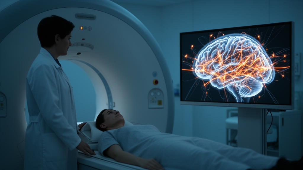
Chronic back pain often begins as a window of opportunity: a subacute phase during which some patients recover while others slide into persistent, life-altering pain. A new study published in Brain Informatics reports that a single resting-state functional MRI scan taken during that subacute phase, combined with machine learning, can distinguish these two trajectories with apparent classification performance well above chance. The work does not offer a clinic-ready test, but it provides one of the more detailed pictures yet of how individualized brain network abnormalities relate to the development of chronic back pain.
Researchers led by Stephan Wunderlich and Sophia Stoecklein analyzed publicly available longitudinal data from the OpenPain database, originally collected at Northwestern University. Their sample comprised 46 patients with subacute back pain, roughly half of them women with a mean age of about 44 years, and 43 healthy controls. Each patient underwent a resting-state fMRI session while still in the subacute stage, and clinical follow-up over the following year allowed the team to classify them as recovered or chronified. Recovery was defined as at least a 20 percent reduction in pain on a visual analog scale one year later; patients who did not reach that threshold were categorized as having progressed toward chronic back pain.
The methodological centerpiece was an individualized measure called the dysconnectivity count. Rather than comparing average connectivity between groups, as most prior studies have done, the researchers compared each patient’s pairwise functional connections against the distribution observed in the healthy control cohort. Any connection deviating by more than three standard deviations from the normative range was flagged as dysconnected, and the dysconnectivity count for each brain region tallied how many of its connections showed such pronounced deviations. This approach, the authors argue, captures patient-specific network abnormalities rather than group-level averages, addressing a long-standing limitation in a field where individual variability has frustrated efforts to build personalized prognostic tools.
Three machine learning models—random forest, elastic net logistic regression, and support vector machine—were then trained to separate future chronic patients from future recoverers based on these regional dysconnectivity counts. Performance was evaluated with repeated random subsampling across 1,000 iterations, with feature selection, training, and weighting performed strictly within each split’s training data. The best result came from a compact set of six brain regions: the mean area under the receiver operating characteristic curve was 0.87, with accuracy of 82 percent, sensitivity of 80 percent, and specificity of 84 percent. A permutation analysis using 1,000 shuffled outcome labels produced a mean AUC of only 0.53, and just one permutation matched or exceeded the observed value, yielding an empirical permutation p-value of 0.002.
The six regions that carried the model were the orbital part of the left inferior frontal gyrus, the opercular part of the right inferior frontal gyrus, the right inferior temporal gyrus, the medial part of the right orbitofrontal gyrus, the right postcentral gyrus, and the right superior frontal gyrus. Each was selected in 82 to 91 percent of training iterations. Dysconnectivity in the inferior temporal gyrus and the two inferior frontal regions was significantly higher in patients who later chronified, driven largely by hyperdysconnectivity—connections exceeding the normal range. In contrast, the orbitofrontal, postcentral, and superior frontal regions showed significantly higher dysconnectivity in patients who recovered, with the medial orbitofrontal region characterized by significant hypodysconnectivity, meaning unusually few connections fell outside the healthy distribution.
The authors interpret this pattern through the lens of prior literature on pain chronification. The inferior frontal and orbitofrontal regions have been implicated in emotion regulation, reward processing, reinforcement learning, and the suppression of intrusive thoughts, while the postcentral gyrus handles sensory integration and the superior frontal gyrus participates in attention and self-referential thinking. Hyperdysconnectivity in the frontal regions of chronic progressors might reflect exaggerated engagement of emotion and reward circuits, whereas altered connectivity in the orbitofrontal and sensory regions of recoverers might reflect protective normalization. Importantly, the researchers are explicit that these psychological constructs—catastrophizing, avoidance, reinforcement learning—were not directly measured in the study, so the mechanistic interpretation remains hypothesis-generating rather than demonstrated.
To translate the six-region pattern into something interpretable, the team built an exploratory composite score weighting each region’s standardized dysconnectivity value by its machine-learning feature importance, with positive contributions from regions more dysconnected in chronic patients and negative contributions from those more dysconnected in recoverers. The score averaged 1.25 in the chronic group versus 0.44 in the recovery group, a highly significant difference. Using a threshold of 0.603 chosen by Youden’s Index, the apparent in-sample positive predictive value was 82.1 percent and the negative predictive value 81 percent. The authors repeatedly caution that these figures, along with likelihood ratios of 5.0 and 0.24, were derived and evaluated in the same cohort and cannot be treated as externally validated performance.
Several sensitivity analyses temper the headline numbers. When the split was changed to a stratified 70/30 train-test design, the AUC dropped to 0.81, which the authors read as evidence that the initial estimate was likely optimistic. Using a more conservative dysconnectivity threshold of 3.5 standard deviations yielded an AUC of 0.84 while retaining all six regions, suggesting the core pattern is robust to threshold choice. Adding age and sex as model inputs actually reduced performance to an AUC of 76 percent, and no demographic variables ranked among the top features. The team also notes that because the 1,000 resampling iterations drew from the same 46 patients, the high feature-selection frequencies reflect stability within overlapping resampling and do not guarantee the same regions would emerge in an independent cohort.
The study’s limitations are candidly laid out. The chronic and recovery groups contained 23 patients each, a small sample prone to statistical noise and optimistic bias. Spinal MRI data were unavailable, so structural spine pathology could not be assessed as a competing explanation. Resting-state connectivity cannot reveal whether hyper- or hypodysconnectivity reflects excitatory or inhibitory neural activity. The atlas used also excluded some regions central to pain chronification in earlier work, including the periaqueductal gray, and the nucleus accumbens and dorsolateral prefrontal cortex—previously highlighted in subacute-to-chronic transition studies—did not emerge among the top features, so the findings may reflect complementary prefrontal alterations rather than a failure to replicate specific earlier pathways.
What the study offers, ultimately, is a proof of concept that individualized dysconnectivity profiling during the subacute phase carries prognostic information about pain trajectory. The authors suggest that, once validated in larger independent cohorts with behavioral measures and spinal imaging, such scans could eventually help identify at-risk patients early and direct them toward preventive interventions. They also point to large cohort evidence that depression and anxiety, rather than spinal imaging findings, are among the strongest predictors of back pain outcomes, arguing that brain-based markers may capture relevant risk where structural spine scans do not. For now, the message is measured: a six-region dysconnectivity signature, an exploratory score, and a clear roadmap of the validation still required before any clinical use.
Subject of Research: Resting-state fMRI dysconnectivity patterns associated with the transition from subacute to chronic back pain
Article Title: Brain dysconnectivity patterns associated with chronic back pain development
Article References: Wunderlich, S., Schulz, E., Ringel, F., Stoecklein, V. M., & Stoecklein, S. (2026). Brain dysconnectivity patterns associated with chronic back pain development. Brain Informatics, 13(1), Article 39. https://doi.org/10.1186/s40708-026-00328-8
Image Credits: AI Generated
DOI: 10.1186/s40708-026-00328-8
Keywords: chronic back pain, resting-state fMRI, functional connectivity, machine learning, dysconnectivity, subacute back pain, pain chronification, brain biomarker, prefrontal cortex, prognostic marker, OpenPain dataset, reinforcement learning
Cite Scienmag News
Cassandra Pierce. (October 29, 2026). Brain Connectivity Patterns May Flag Which Back Pain Patients Will Chronicify. Scienmag. https://scienmag.com/brain-connectivity-patterns-may-flag-which-back-pain-patients-will-chronicify/
Cassandra Pierce. "Brain Connectivity Patterns May Flag Which Back Pain Patients Will Chronicify." Scienmag, 29 October 2026, https://scienmag.com/brain-connectivity-patterns-may-flag-which-back-pain-patients-will-chronicify/. Accessed 30 October 2026.
Cassandra Pierce. "Brain Connectivity Patterns May Flag Which Back Pain Patients Will Chronicify." Scienmag. October 29, 2026. https://scienmag.com/brain-connectivity-patterns-may-flag-which-back-pain-patients-will-chronicify/