


Anxiety in adolescence has long been treated as a diffuse, hard-to-pin-down phenomenon, a cluster of symptoms that emerges as the teenage brain undergoes its most dramatic period of reorganization. Now a large-scale neuroimaging study from Boston Children’s Hospital offers something the field has lacked: a specific, measurable brain circuit whose altered organization tracks with anxiety in young people, along with evidence that the same alterations carry cognitive costs that extend well beyond mood. The research, published in the journal Imaging Neuroscience, draws on data from nearly 3,500 adolescents and points toward a future in which psychiatric care for youth could be tailored to the individual wiring of the brain.
Adolescence is widely recognized as a sensitive window for both brain development and mental health. Many psychiatric conditions, including anxiety disorders, make their first appearance during these years, yet the precise relationship between the developing brain’s changing architecture and the emergence of symptoms has remained incompletely understood. The stakes are considerable: anxiety disorders, including generalized anxiety disorder and social anxiety disorder, affect more than 30 percent of teenagers in the United States, making them among the most common mental health conditions of youth. Understanding which neural systems are involved, and how early their signatures become detectable, could reshape when and how clinicians intervene.
The study was led by Caterina Stamoulis, PhD, Principal Investigator in the Computational Neuroscience Laboratory at Boston Children’s Hospital. Her team took advantage of the Adolescent Brain Cognitive Development Study, or ABCD, the largest long-term study of brain development and child health ever conducted in the United States. By examining associations between the structural characteristics of brain regions, the organization of the circuits they form, and measures of anxiety across a sample of nearly 3,500 youth, the researchers sought to move beyond broad correlations between whole-brain measures and behavior and instead identify specific networks implicated in the disorder.
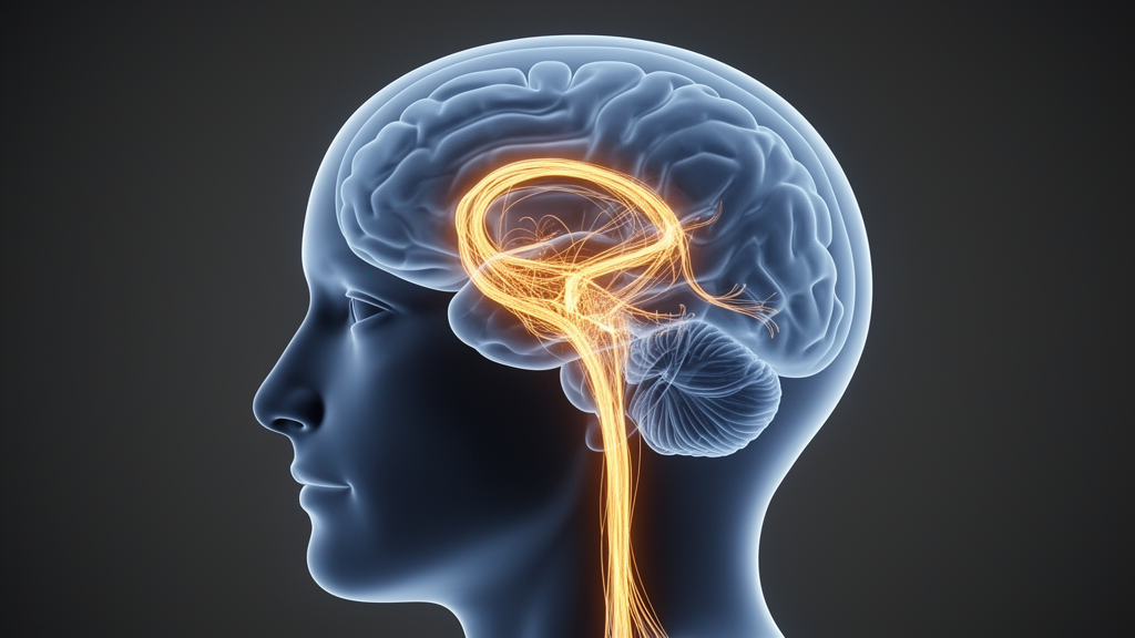
What they found was a circuit composed of three structures: the cerebellum, the amygdala, and the thalamus. Each of these plays a distinctive role in brain function. The amygdala, buried deep in the temporal lobes, is central to processing threat and emotional salience. The thalamus acts as a major relay station, gating the flow of sensory and other information to the cortex. The cerebellum, long associated with motor coordination, is now understood to participate in a wide range of cognitive processes and to exert regulatory roles over brain function more broadly. In youth with anxiety, the organization of this three-node circuit was altered, and the strength of its connections was reduced.
Those alterations were not merely correlates of a diagnostic label. The researchers report that weaker and less well-organized connections within the circuit were associated with higher levels of anxiety and with a greater likelihood of related disorders. In other words, the circuit’s properties varied in a graded fashion with symptom severity, a pattern consistent with the idea that the circuit is functionally involved in the mechanisms of anxiety rather than simply marking its presence. At the same time, the study identified a protective counterpart: the efficiency and resilience, or robustness, of other brain networks were associated with lower risk of social and generalized anxiety disorders. The developing brain, in this view, is not a single system tipping toward or away from illness but a collection of networks whose competing properties shape mental health outcomes.
The findings did not stop at circuit organization. The team also identified structural alterations in the brains of youth with anxiety, and crucially, they found that these structural and circuit alterations were associated with reduced performance in a specific cognitive domain: the ability to perceive, process, and integrate information about spatial locations and the relationships between objects and shapes. This capacity, known as visuospatial processing, is far from an abstract laboratory construct. It underpins everyday tasks ranging from navigating spaces and driving a car to assembling furniture and tying shoes. The implication is that anxiety in adolescence may be accompanied by measurable deficits in cognitive skills that young people rely on daily, a connection that has rarely been drawn so explicitly in previous work.
Perhaps the most consequential finding concerns timing. The anxiety-related brain alterations were already measurable at ages 11 to 12, a point at which many of the study participants had not yet developed clinically significant anxiety symptoms. This early detectability reframes the clinical picture. If the neural signatures of vulnerability are present before the full syndrome emerges, then the window for preventive intervention opens years earlier than current practice typically assumes. Waiting for symptoms to become impairing may mean intervening after the brain’s developmental trajectory has already been shaped, whereas earlier action could minimize long-term cognitive deficits and mental health problems.
Stamoulis sees in these results an argument for precision medicine applied to psychiatry. The identification of specific circuits implicated in anxiety, she suggests, provides concrete targets for interventions designed to improve both short-term and longer-term mental health outcomes in youth. Rather than treating adolescent anxiety as a homogeneous condition addressed with one-size-fits-all approaches, clinicians could in principle use neuroimaging markers to identify which young people show alterations in this cerebellar-amygdala-thalamic circuit and tailor treatments accordingly. Such precision psychiatry, she notes, is likely to play an important role in future mental health care, though the study itself establishes associations rather than demonstrating that altering the circuit directly changes anxiety outcomes.
The methodological strength of the work lies in its scale and its analytical approach. Studies of adolescent brain development have often been limited by small samples, which make it difficult to distinguish genuine neural signatures from statistical noise. By drawing on the ABCD cohort, the Boston Children’s team could examine circuit properties across a large and diverse population of youth, increasing confidence that the associations they observed reflect robust features of the developing brain rather than artifacts of a particular group. The combination of structural measures, network organization metrics, cognitive testing, and clinical assessments of anxiety allowed the researchers to connect levels of analysis that are usually studied in isolation.
The broader significance of the study extends beyond anxiety itself. By showing that a defined circuit involving the cerebellum, amygdala, and thalamus carries both emotional and cognitive consequences, the work reinforces a growing recognition that mental health and cognition are intertwined at the level of brain networks. It also highlights the value of large longitudinal resources like ABCD, which allow scientists to track how brain organization and behavior co-evolve across adolescence. For the millions of teenagers affected by anxiety, and for the clinicians who treat them, the study offers a concrete starting point: a map of where to look, a timeline for when to look, and a rationale for acting before symptoms take hold. As precision interventions are developed and tested, the circuit identified by Stamoulis and her colleagues may well become one of the first neural targets in a new era of youth mental health care.
Subject of Research: Neural circuit alterations associated with adolescent anxiety identified through large-scale neuroimaging of the developing brain
Article Title: Researchers identify specific brain circuits associated with anxiety in adolescents
Article References: Researchers identify specific brain circuits associated with anxiety in adolescents. (n.d.). Original publication
Image Credits: AI Generated
DOI: Not provided
Keywords: adolescent anxiety, brain circuits, cerebellum, amygdala, thalamus, ABCD study, neuroimaging, precision psychiatry, visuospatial processing, brain development, Boston Children's Hospital, Imaging Neuroscience
Cite Scienmag News
Glenn Wilkins. (October 9, 2026). Brain Circuit Linking Cerebellum, Amygdala and Thalamus Tied to Teen Anxiety. Scienmag. https://scienmag.com/brain-circuit-linking-cerebellum-amygdala-and-thalamus-tied-to-teen-anxiety/
Glenn Wilkins. "Brain Circuit Linking Cerebellum, Amygdala and Thalamus Tied to Teen Anxiety." Scienmag, 9 October 2026, https://scienmag.com/brain-circuit-linking-cerebellum-amygdala-and-thalamus-tied-to-teen-anxiety/. Accessed 9 October 2026.
Glenn Wilkins. "Brain Circuit Linking Cerebellum, Amygdala and Thalamus Tied to Teen Anxiety." Scienmag. October 9, 2026. https://scienmag.com/brain-circuit-linking-cerebellum-amygdala-and-thalamus-tied-to-teen-anxiety/


