


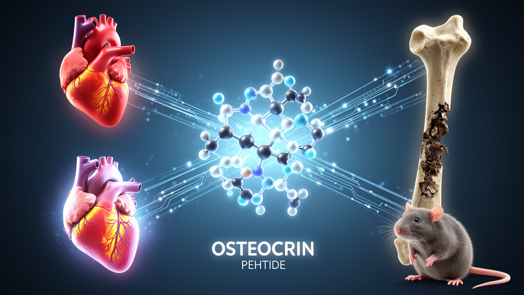
A little-known hormone made in bone has emerged as an unexpected signal of heart disease, according to a new translational study that followed both sick dogs and laboratory rats. Researchers report that circulating concentrations of osteocrin, a secreted peptide best known for its roles in bone biology and natriuretic peptide regulation, changed in step with the severity of myxomatous mitral valve disease in a cohort of 47 dogs and rose in parallel with bone deterioration in a rat model of pressure-overload cardiac remodeling. The work, published in Frontiers in Veterinary Science, positions osteocrin as a candidate biomarker at the interface between cardiovascular and skeletal remodeling, while stopping short of any claim that the peptide causes disease.
Myxomatous mitral valve disease is the most common acquired cardiac condition in dogs, marked by progressive degeneration of the mitral valve leaflets, leaking of blood backward through the valve, and gradual remodeling of the heart chambers that can end in congestive heart failure. Veterinarians grade its severity with the American College of Veterinary Internal Medicine staging system, from stage B1 through B2 to the advanced C/D categories. Increasingly, heart failure is understood as a systemic disorder, and human studies have linked cardiovascular disease with reduced bone mineral density and elevated fracture risk, raising the question of whether a single circulating molecule might reflect both processes at once.
Osteocrin, also called musclin, is a secreted peptide produced in bone and skeletal muscle that regulates the clearance of natriuretic peptides through interactions with natriuretic peptide receptor-C, a receptor that controls tissue availability of these heart-derived signaling molecules. Previous experimental work has also detected osteocrin in cardiac tissue and implicated it in cardiovascular stress responses and cardioprotection in models of cardiac injury. What remained unclear was whether circulating osteocrin concentrations track disease severity in naturally occurring cardiovascular disease and whether they relate to skeletal remodeling in a complementary experimental setting. The Japanese research team, led by Mitsuhiro Isaka of Rakuno Gakuen University, hypothesized that the peptide would be associated with both.
To test this, the investigators combined two very different study arms. Between April 2019 and May 2020, they enrolled 47 client-owned dogs at the Yokohama Yamate Dog and Cat Medical Center and the Rakuno Gakuen University Veterinary Teaching Hospital: 31 dogs with myxomatous mitral valve disease, classified as stage B1 (10 dogs), B2 (14) or C/D (7), and 16 clinically healthy controls. Each dog underwent physical examination, thoracic radiography and echocardiography, with measurements including the left atrial-to-aorta ratio, left ventricular internal diameters, fractional shortening, ejection fraction and vertebral heart size. Serum osteocrin concentrations were measured with enzyme-linked immunosorbent assays run in duplicate.
The canine results revealed a striking stage-dependent pattern. Dogs with stage B1 disease had significantly lower serum osteocrin concentrations than healthy controls and stage B2 dogs, whereas dogs in the advanced C/D stages had significantly higher concentrations than healthy controls and both earlier stages. Osteocrin also correlated positively with several echocardiographic indices of cardiac remodeling: the left atrial-to-aorta ratio (Spearman’s rs = 0.61), left ventricular internal diameter in diastole (rs = 0.67), fractional shortening (rs = 0.50) and vertebral heart size (rs = 0.41), all statistically significant, while no significant correlation appeared with ejection fraction. In a multivariable linear regression including 46 dogs with complete data, ACVIM stage remained significantly associated with osteocrin after adjustment for age and body weight, with stage B1 showing a lower adjusted concentration (β = −0.759) and stages B2 and C/D showing higher values (β = 0.321 and 1.027 respectively). The model explained 74.0 percent of the variability in serum osteocrin.
Because Chihuahuas were disproportionately represented among the diseased dogs, the team ran a sensitivity analysis adding breed as a covariate. Chihuahua status itself was not independently associated with osteocrin concentrations, and the associations for stages B1 and C/D remained significant, though the stage B2 association was attenuated and no longer reached statistical significance. The authors caution that residual breed-related confounding cannot be completely excluded, and they emphasize that the observational design precludes any inference of causality. They also note that the lower osteocrin values in stage B1 remain unexplained and may reflect biological heterogeneity, underscoring the need for validation in larger longitudinal cohorts.
The second arm of the study turned to an experimental model. Six-week-old female Wistar rats were assigned to abdominal aortic constriction, an established method of inducing pressure-overload cardiac remodeling, or to sham surgery, with six animals per group followed for 21 days. Female animals were chosen because osteoporosis is more prevalent in women, aligning the design with the study’s focus on cardiovascular-skeletal interactions. Histology at the endpoint confirmed structural remodeling: left ventricular wall thickness was greater in constricted rats (2,281.3 versus 1,879.3 micrometers, P = 0.0245), as was interventricular septal thickness (2,625.6 versus 2,084.4 micrometers, P = 0.0331). The researchers deliberately avoid calling this a heart failure model, noting that trans-stenotic pressure gradients and left ventricular pressures were not directly measured, so the magnitude of the hemodynamic load cannot be quantified.
The biomarker dynamics in rats proved telling. Two-way repeated-measures analysis of variance showed significant effects of group, time and their interaction for osteocrin, with postoperative concentrations rising significantly within the pressure-overload group and ending significantly higher than in controls. By contrast, NT-proBNP, the well-established clinical heart failure biomarker, showed no significant pressure-overload-specific increase over the 21-day window; only a decline in the sham group reached significance. The authors suggest this may reflect the early remodeling stage captured by the short follow-up, but they stress that whether osteocrin responds earlier than NT-proBNP during cardiovascular remodeling cannot be determined from the present data and requires longitudinal study with detailed cardiac phenotyping.
The skeletal side of the rat experiment added a further layer. Dual-energy X-ray absorptiometry revealed significantly reduced bone mineral density in the femurs of pressure-overloaded rats, and three-point bending tests showed significant reductions in both maximum load and stiffness, each at P < 0.001. In pooled analyses across all 12 animals, circulating osteocrin was strongly inversely correlated with bone mineral density (rs = −0.944) and stiffness (rs = −0.854), while NT-proBNP showed no significant association with any skeletal parameter. However, the authors are unusually candid about the limits of these correlations: when analyzed separately within each group, the osteocrin-bone associations did not reach statistical significance, indicating that the strong pooled relationships were substantially driven by the separation between the two groups rather than a continuous dose-response relationship at the individual-animal level. Immunohistochemistry added a spatial clue, localizing osteocrin immunoreactivity predominantly to cells lining the atrioventricular valvular surface, with no detectable signal in myocardial fibers, though localization alone cannot establish the source of the circulating peptide.
The study carries a long list of acknowledged limitations. The clinical design was cross-sectional and observational; NT-proBNP was not measured in the dogs, preventing direct comparison with established cardiac biomarkers; echocardiographic data were incomplete for several variables, with missingness concentrated among healthy dogs; only female rats were studied, limiting generalizability to males; the exploratory correlation analyses were not adjusted for multiple testing; and chronic kidney disease, documented in 7 of 47 dogs, could not be classified by azotemia status because renal biochemistry was unavailable, leaving residual confounding possible. Even so, the convergent picture from a natural disease cohort and an experimental model supports further investigation of osteocrin as a candidate marker of coordinated cardiovascular and skeletal remodeling. Whether the peptide actively participates in heart-bone communication or merely reflects it remains an open question that future longitudinal and mechanistic studies will need to answer.
Subject of Research: Circulating osteocrin as a candidate biomarker linking cardiac remodeling in canine mitral valve disease with skeletal alterations in experimental pressure overload
Article Title: Circulating osteocrin is associated with cardiac remodeling in canine myxomatous mitral valve disease and skeletal alterations in a rat pressure-overload model
Article References: Komatsu, M., Udagawa, H., Araki, R., Hamamoto, Y., Nam, E., Okamoto, M., & Isaka, M. (2026). Circulating osteocrin is associated with cardiac remodeling in canine myxomatous mitral valve disease and skeletal alterations in a rat pressure-overload model. Frontiers in Veterinary Science, 13, Article 1975572. https://doi.org/10.3389/fvets.2026.1975572
Image Credits: AI Generated
DOI: 10.3389/fvets.2026.1975572
Keywords: osteocrin, myxomatous mitral valve disease, cardiac remodeling, natriuretic peptides, pressure overload, bone mineral density, NT-proBNP, canine cardiology, rat model, heart-bone axis, biomarker, veterinary medicine
Cite Scienmag News
William Thompson. (October 24, 2026). Bone-Derived Peptide Osteocrin Tracks Heart Remodeling in Dogs and Bone Loss in Rats. Scienmag. https://scienmag.com/bone-derived-peptide-osteocrin-tracks-heart-remodeling-in-dogs-and-bone-loss-in-rats/
William Thompson. "Bone-Derived Peptide Osteocrin Tracks Heart Remodeling in Dogs and Bone Loss in Rats." Scienmag, 24 October 2026, https://scienmag.com/bone-derived-peptide-osteocrin-tracks-heart-remodeling-in-dogs-and-bone-loss-in-rats/. Accessed 24 October 2026.
William Thompson. "Bone-Derived Peptide Osteocrin Tracks Heart Remodeling in Dogs and Bone Loss in Rats." Scienmag. October 24, 2026. https://scienmag.com/bone-derived-peptide-osteocrin-tracks-heart-remodeling-in-dogs-and-bone-loss-in-rats/


