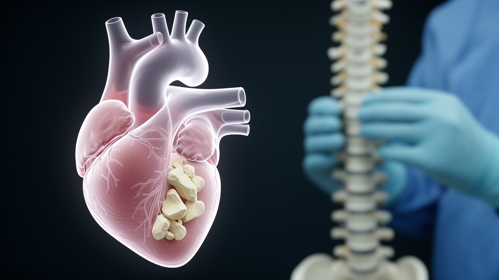
A 68-year-old woman developed sudden abdominal distension, chest tightness, and shortness of breath just five days after undergoing percutaneous kyphoplasty, a minimally invasive spine procedure in which balloon-inflated cavities are filled with bone cement. When clinicians investigated, they found that the trouble did not come from the cement injected during that operation. Instead, imaging pointed to cement that had leaked from a vertebra treated at another hospital three months earlier, traveled through the venous system, and lodged in her heart. The case, published as a case report in Discover Medicine, describes how the embolus was managed conservatively after the patient declined surgical removal, and how she returned to normal life at her three-month and six-month follow-up appointments.
The patient had first been admitted for one day of chest and back pain caused by trauma and was diagnosed with a compression fracture of the twelfth thoracic vertebral body, or T12. Her history revealed that three months before this admission she had undergone kyphoplasty at another hospital for traumatic compression fractures of the seventh and tenth thoracic vertebrae and the first lumbar vertebra. Postoperative X-rays taken at that time had already shown that bone cement had leaked into the heart via the inferior vena cava, yet the patient reported no obvious discomfort then. The authors note that a definitive causal relationship between the old cement leak and her new symptoms could not be established from the available evidence, although preoperative and postoperative imaging suggested the symptoms were possibly related to cement leakage from the T7 vertebra.
At the current hospital, after completing preoperative examinations, surgeons performed percutaneous kyphoplasty under local anesthesia. They injected 3 milliliters of polymethylmethacrylate, a standard acrylic bone cement, into bilateral regions of the T12 vertebral body under C-arm fluoroscopy. Intraoperative fluoroscopy confirmed that the cement was evenly distributed, that the height of the vertebral body had been recovered, and that the cement was properly positioned within the vertebral body without leakage or spinal canal invasion. On that basis, the operation itself appeared technically uneventful.
The crisis emerged on the fifth postoperative day, when the patient suddenly developed abdominal distension, chest tightness, and dyspnea at night. Physical examination revealed obvious abdominal distension without muscular tension or board-like rigidity. Oxygen inhalation and vital sign monitoring were initiated immediately, showing a blood oxygen saturation of 90 percent. An urgent general surgery consultation was arranged, and blood gas analysis showed decreases in both the partial pressure of oxygen and the partial pressure of carbon dioxide, with a PaO2 of 78 mmHg and a PaCO2 of 33 mmHg. She was transferred to the intensive care unit for symptomatic supportive treatment that included heparin anticoagulation.
Imaging then clarified what was happening. An archived paper-based echocardiography report described a 26 by 20 millimeter hyperechoic irregular mass in the right heart. The report’s authors are candid about the limitations of that document: it contained non-standard and internally inconsistent anatomical terminology, such as a reference to a “left posterior lobe of the pulmonary valve,” which could not be reliably reconciled with a right-heart location, and the original electronic echocardiographic images were unavailable for review. Color Doppler echocardiography raised suspicion of a bone cement embolus in the right heart, rather than specifically in the right atrium, with risks of cardiac perforation and embolus shedding. Chest X-ray confirmed the presence of a bone cement embolus in the right heart.
After one day in the intensive care unit the patient’s condition improved significantly, with stable general status and stable vital signs, and she was transferred back to the general ward. A cardiac surgery consultation recommended surgical removal of the intracardiac cement embolus at an appropriate time. However, citing economic and psychological factors, the patient refused to undergo embolectomy. Her condition remained stable, her pain resolved one week after surgery, and she was discharged once she could ambulate. She was continued on warfarin anticoagulation for six months, a decision the authors describe as individualized based on limited case-based evidence, intended to reduce the risk of thrombus forming on the surface of the cement embolus and of subsequent thromboembolic complications. At the three-month and six-month follow-ups she had returned to normal life with no subjective discomfort, and repeated imaging consistently showed the residual embolus remained stable.
The report places this case in a broader clinical context. Percutaneous kyphoplasty is widely used to treat conditions including osteoporotic vertebral compression fractures and spinal cord metastases. Bone cement leakage is its most common complication, with an incidence of approximately 23.3 percent after kyphoplasty, significantly lower than the 37.3 percent reported after percutaneous vertebroplasty. Yet it is extremely rare for leaked cement to enter the heart through the venous system and cause cardiac and pulmonary embolism, with only very few clinical cases reported. A retrospective study cited in the paper found that approximately 8.3 percent of patients were diagnosed with intracardiac bone cement embolism, yet only six of these patients presented with obvious symptoms, meaning most affected patients were asymptomatic, a situation the authors warn is often overlooked by clinicians. Once severe adverse events occur, they can lead to fatal outcomes such as cardiac perforation and cardiopulmonary failure.
The pathological route is relatively well understood. Leaked cement primarily enters the inferior vena cava via paravertebral veins. Most cement emboli then travel with the bloodstream into the right atrium and onward into the pulmonary artery, triggering pulmonary embolism. In a small number of cases, if the embolus is too large or has an irregular, sharp shape, it cannot pass through the heart valves, which may result in cardiac perforation or valve injury. Unlike thromboemboli, bone cement emboli can be clearly visualized on X-ray because cement is denser than lung parenchyma. The authors also detail factors that influence leakage, including the volume and viscosity of injected cement, the integrity of the posterior wall of the fractured vertebra, and the operator’s experience. Higher-viscosity cement can stabilize compressed bone while minimizing leakage risk, and injection is generally performed during the drawing stage, when the cement has a toothpaste-like consistency. Unilateral and bilateral puncture approaches show no significant difference in leakage rates, and one cited analysis identified 13.68 percent as the optimal ratio of puncture-side cement volume to vertebral body volume, above which the risk of paravertebral vein leakage increases significantly.
Management of such emboli remains unsettled. The authors distinguish between two goals: preventing thrombus formation on the surface of the cement and preventing migration of the cement itself. Anticoagulation addresses only the former; it cannot prevent mechanical migration of the solid cement fragment. Published suggestions hold that asymptomatic cardiac cement emboli can be managed conservatively with anticoagulation until the embolus becomes epithelialized, while symptomatic patients should receive interventional removal, preferring percutaneous surgery when cement sits in the right atrium and open surgery when it sits in the right ventricle. The report stresses that these recommendations rest on very limited literature and thus have limited clinical reference value. The authors speculate that patients with pericardial hematocele, obvious right ventricular perforation, hemodynamic disorder, or cardiopulmonary failure caused by embolism have clear indications for surgical intervention, but for patients without severe symptoms, especially elderly patients with comorbidities, the risks and benefits of surgery versus conservative treatment require careful weighing.
The case’s limitations are acknowledged directly: the patient declined further surgical intervention, six-month follow-up data were available, but longer-term follow-up is lacking and the long-term outcomes of conservative management remain unclear. The authors recommend routine postoperative chest X-ray after kyphoplasty, with echocardiography and chest CT arranged when necessary, to identify asymptomatic patients with pulmonary or intracardiac cement embolism before severe cardiopulmonary failure develops. They propose close regular follow-up, at least once every three months during the first year after diagnosis and roughly every six months to one year thereafter, while noting that the optimal schedule and long-term outcomes still require further investigation. Routine cement injection under C-arm fluoroscopy monitoring, they conclude, can reduce the risk of leakage in the first place.
Subject of Research: A case report of intracardiac bone cement embolism following percutaneous kyphoplasty
Article Title: A case report of intracardiac cement embolism after percutaneous kyphoplasty
Article References: Shu, Q. (2026). A case report of intracardiac cement embolism after percutaneous kyphoplasty. Discover Medicine, 3(1), Article 159. https://doi.org/10.1007/s44337-026-00726-1
Image Credits: AI Generated
DOI: 10.1007/s44337-026-00726-1
Keywords: percutaneous kyphoplasty, bone cement, intracardiac embolism, cement leakage, case report, polymethylmethacrylate, spinal fracture, echocardiography, anticoagulation, conservative management, vertebroplasty, complications
Cite Scienmag News
Ophelia Keating. (October 28, 2026). Bone Cement Leaks Into a Patient’s Heart After Spine Surgery, in Rare Case Report. Scienmag. https://scienmag.com/bone-cement-leaks-into-a-patients-heart-after-spine-surgery-in-rare-case-report/
Ophelia Keating. "Bone Cement Leaks Into a Patient’s Heart After Spine Surgery, in Rare Case Report." Scienmag, 28 October 2026, https://scienmag.com/bone-cement-leaks-into-a-patients-heart-after-spine-surgery-in-rare-case-report/. Accessed 28 October 2026.
Ophelia Keating. "Bone Cement Leaks Into a Patient’s Heart After Spine Surgery, in Rare Case Report." Scienmag. October 28, 2026. https://scienmag.com/bone-cement-leaks-into-a-patients-heart-after-spine-surgery-in-rare-case-report/