


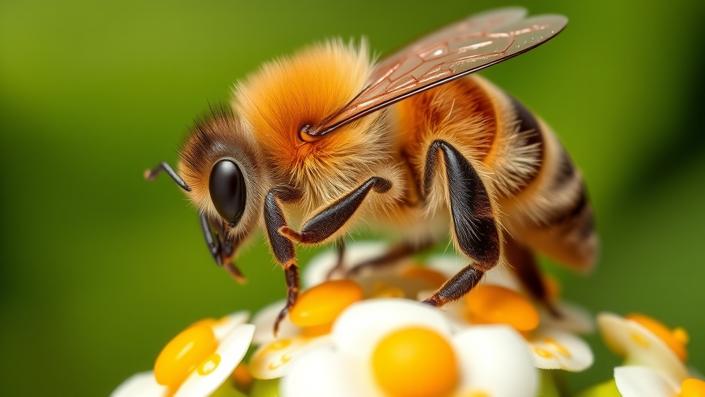
Propolis, the resinous substance that honeybees use to seal and sterilize their hives, may hold unexpected therapeutic value far beyond the hive walls. A new study published in the journal 3 Biotech reports that propolis effectively protected mice from intestinal injury triggered by lipopolysaccharide, or LPS, a toxic component of the outer membrane of Gram-negative bacteria that is a well-known driver of inflammation and tissue damage in the gut. The research, led by Muyuan Niu, Jia Le, Shiying Liao and Kun Li from Nanjing Agricultural University and Hubei University of Arts and Science, provides a detailed mechanistic picture of how this natural bee product simultaneously calms inflammation, bolsters antioxidant defenses, strengthens the physical intestinal barrier and reshapes the composition and function of the gut microbiota.
Diarrhea-causing bacteria remain one of the most common causes of intestinal inflammation and injury worldwide, and the search for interventions that go beyond antibiotics has intensified as concerns about drug resistance grow. Propolis has long attracted attention because it is packed with bioactive compounds that exhibit anti-inflammatory and antioxidant activities in laboratory settings. Yet, as the authors note, its role in maintaining the integrity of the intestinal barrier and in modulating the trillions of microbes that inhabit the gut has remained insufficiently understood. The new study was designed specifically to close that gap by testing propolis in a controlled model of acute intestinal damage.
The experimental setup was straightforward but rigorous. Thirty Institute of Cancer Research mice were randomly divided into three groups: a normal control group, an LPS-treated model group, and a propolis treatment group. Mice in the model group received an intraperitoneal injection of LPS at a dose of 10 milligrams per kilogram of body weight, which reliably induces systemic inflammatory stress and measurable injury to the small intestine. Mice in the treatment group received propolis at 75 milligrams per kilogram by the same route. By comparing these groups across a battery of molecular, histological and microbiological endpoints, the team could isolate the specific protective effects attributable to propolis.
The first and most visible sign of protection came from body weight. LPS injection typically causes mice to lose weight as inflammation takes its metabolic toll, and the animals in the model group followed that expected pattern. In contrast, the mice that received propolis lost significantly less weight, an early indication that the treatment was buffering the physiological impact of the LPS challenge. The protective effect was even more striking under the microscope. Examination of the jejunum, the middle section of the small intestine where nutrient absorption is concentrated, revealed that propolis improved intestinal morphology by increasing villus height and improving the ratio of villus height to crypt depth, both classic indicators of a healthier, more functional intestinal lining.
Beneath these structural improvements, the researchers documented a profound shift in the inflammatory chemistry of the treated animals. LPS drives the release of proinflammatory cytokines, signaling molecules that recruit immune cells and amplify tissue damage. In the propolis-treated mice, the levels of the key proinflammatory mediators IL-1β, TNF-α and IL-6 were significantly lowered, while the level of IL-10, an anti-inflammatory cytokine that helps resolve inflammation and restore tissue homeostasis, rose at the same time. This dual action, suppressing the inflammatory cascade while enhancing its natural counterweights, suggests that propolis does not simply blunt immune signaling wholesale but actively rebalances it.
Oxidative stress is the other half of the LPS damage equation. When inflammatory pathways are triggered, cells generate reactive oxygen species that attack lipids, proteins and DNA, and the extent of this damage is commonly measured by levels of malondialdehyde, or MDA, a marker of lipid peroxidation. In the propolis-treated mice, MDA levels fell, while the activities of the antioxidant enzymes superoxide dismutase, total antioxidant capacity, and glutathione peroxidase all increased. Taken together, these measurements indicate that propolis fortified the animals’ endogenous antioxidant defenses, allowing their tissues to neutralize the reactive molecules that would otherwise compound the injury caused by inflammation.
Perhaps the most clinically significant finding concerns the intestinal barrier itself. The lining of the gut is held together by tight junctions, protein complexes that seal the gaps between epithelial cells and prevent bacteria and toxins from crossing into the bloodstream. Three sentinel proteins, Claudin-1, ZO-1 and Occludin, are widely used as markers of barrier integrity. In the propolis-treated mice, the gene expression and protein levels of all three markers were significantly upregulated, indicating that the tight junctions had been reinforced at both the transcriptional and translational levels. A stronger barrier means less bacterial translocation, less endotoxin leakage and a lower risk of the systemic inflammatory spread that makes intestinal injury so dangerous.
To explore the effects of propolis on the gut microbiota, the team turned to high-throughput 16S rRNA sequencing, generating more than 24 million raw reads and nearly four million filtered reads from the samples. The sequencing data showed that propolis increased the diversity of gut microbes, a hallmark of a resilient and healthy microbial community, and altered the abundance of beneficial bacterial taxa in ways associated with improved gut function. Beyond simply shifting which species were present, propolis enhanced the network of relationships among the microbes, suggesting a more connected and stable ecological community capable of resisting disruption.
The functional consequences of these microbial changes were assessed through predictive metagenomic analysis using MetaCyc and KEGG pathways. The results indicated that propolis restored metabolic functions in the gut microbiome that had been disturbed by the LPS challenge. This is an important dimension of the findings because microbial metabolism influences everything from short-chain fatty acid production to immune signaling, and a microbiome that is both taxonomically diverse and metabolically active is far better equipped to support the host during inflammatory stress. The convergence of structural, immunological, antioxidant, barrier and microbial evidence paints a coherent picture of a compound that acts on multiple fronts at once.
The authors conclude that propolis was effective at preventing intestinal damage caused by LPS through its combined anti-inflammatory, antioxidant, barrier-maintaining and microbiota-balancing actions, and they propose that it should be considered a promising natural therapeutic agent for preventing and treating intestinal inflammation and injury. The work was supported by the Science and Technology Planning Project of Qamdo City, Xizang, and all raw sequencing data from the animals was deposited in the NCBI Sequence Read Archive under accession number PRJNA1400253, allowing other researchers to verify and extend the findings. While the study was conducted in mice and used intraperitoneal administration, the breadth and internal consistency of the results add substantial weight to the growing body of evidence that bee-derived natural products deserve a serious place in the search for safer gut-protective therapies. Whether propolis can deliver the same multi-layered protection in humans, and at what dose and route of administration, will be the critical questions for the next generation of studies.
Subject of Research: Protective effects of propolis against LPS-induced intestinal injury in mice
Article Title: Propolis alleviated intestine injury in LPS treated mice by accommodating inflammation, oxidation resistance, intestinal barrier and microbiota
Article References: Niu, M., Le, J., Liao, S., & Li, K. (2026). Propolis alleviated intestine injury in LPS treated mice by accommodating inflammation, oxidation resistance, intestinal barrier and microbiota. 3 Biotech, 16(10), Article 440. https://doi.org/10.1007/s13205-026-05075-5
Image Credits: AI Generated
DOI: 10.1007/s13205-026-05075-5
Keywords: propolis, lipopolysaccharide, gut microbiota, intestinal barrier, inflammation, oxidative stress, ICR mice, cytokines, tight junctions, 16S rRNA sequencing, intestinal injury, alleviated
Cite Scienmag News
Gregory Coleman. (September 20, 2026). Bee Propolis Shields the Gut From Inflammatory Damage, Mouse Study Finds. Scienmag. https://scienmag.com/bee-propolis-shields-the-gut-from-inflammatory-damage-mouse-study-finds/
Gregory Coleman. "Bee Propolis Shields the Gut From Inflammatory Damage, Mouse Study Finds." Scienmag, 20 September 2026, https://scienmag.com/bee-propolis-shields-the-gut-from-inflammatory-damage-mouse-study-finds/. Accessed 4 October 2026.
Gregory Coleman. "Bee Propolis Shields the Gut From Inflammatory Damage, Mouse Study Finds." Scienmag. September 20, 2026. https://scienmag.com/bee-propolis-shields-the-gut-from-inflammatory-damage-mouse-study-finds/


