


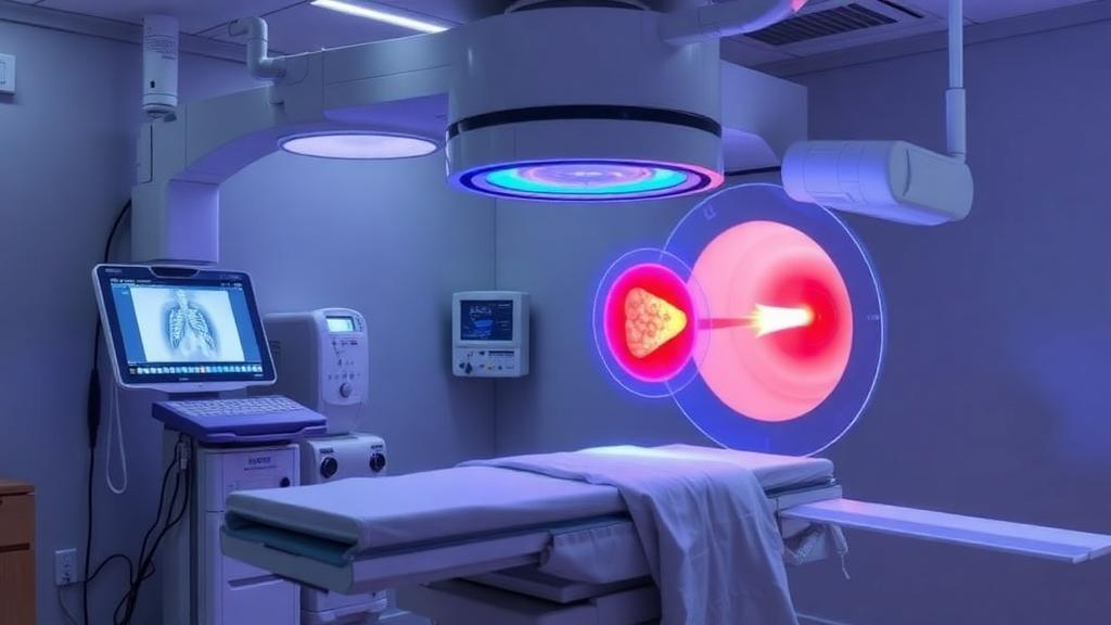
For patients with liver tumors, one of the most technically impressive weapons in the interventional radiology arsenal has long carried an inconvenient requirement: general anesthesia. CT-guided high-dose-rate brachytherapy, a radioablative technique in which a tiny iridium-192 radiation source is steered directly into a tumor through temporarily placed catheters, has demonstrated strikingly good local tumor control in previous clinical studies, including evidence that it can outperform transarterial chemoembolization in survival outcomes for treatment-naive patients with unresectable hepatocellular carcinoma. But the question of how patients should be sedated during the procedure has remained unresolved, with practices varying widely between institutions. Now a prospective study from Charité – Universitätsmedizin Berlin offers a compelling answer: the entire procedure can be done safely and comfortably with the patient awake, using only conscious sedation with analgesia administered by the interventional radiology team itself.
The study, published in CVIR Oncology as an open-access short communication, enrolled 97 patients with primary or secondary liver malignancies, of whom 74 completed the final analysis. All participants were treated under conscious sedation with analgesia rather than general anesthesia, and all were interviewed systematically about their pain experience at three distinct time points: during catheter placement under CT fluoroscopy, at the time of radiation delivery when the catheters were removed, and later the same evening on the ward. Pain was scored on the standard numeric rating scale from zero, meaning no pain, to ten, meaning maximum pain. The researchers also tracked age, sex, the number of lesions and catheters, prior treatments, tumor volume, tumor location, and complications classified according to Society of Interventional Radiology guidelines.
The technical choreography of the procedure helps explain why the anesthesia question matters so much. Unlike thermal ablation, where a probe is inserted and the tumor is burned or frozen in a single session, CT-guided HDR brachytherapy unfolds in two stages separated by a change of location. First, under CT fluoroscopic guidance, the interventionalist percutaneously places brachytherapy applicators into and around the tumor while the patient receives intravenous midazolam and fentanyl alongside local lidocaine anesthesia at the puncture site. A second physician stands by throughout, monitoring vital signs and titrating medication. Once the catheters are positioned and the planning scan is complete, the puncture sites are sterilely covered and the patient is escorted to a dedicated radiation operating room, where the iridium-192 source delivers its dose. Only after irradiation are the catheters removed, creating a second moment of potential discomfort, and the patient is then observed for roughly an hour before transfer to the regular ward.
The pain data that emerged were strikingly reassuring. At catheter placement, the moment many clinicians would expect to hurt most, 74.5 percent of patients rated their pain as low, in the zero-to-two range, while 19.0 percent reported moderate pain and only 6.5 percent severe pain. At the time of radiation and catheter removal, the picture improved further: 84.0 percent reported low pain, 11.0 percent moderate, and 5.5 percent severe. On the ward afterward, 79.5 percent reported low pain, 16.0 percent moderate, and 4.0 percent severe. The median doses of sedation were modest, 112.5 micrograms of fentanyl and 2 milligrams of midazolam, underscoring how little pharmacological support the procedure actually demands when performed with careful local anesthesia and image guidance.
Two patterns within the data carry practical weight for clinicians. Women reported significantly higher pain at the time of irradiation, with a p-value below 0.001, and the number of treated lesions was associated with significantly higher pain levels at two of the three measured time points, with p below 0.05. No other parameter, including age, tumor volume, tumor location, or the number of prior treatments, showed a significant relationship with pain perception. The authors suggest that these findings point to patient groups, women and those with multiple lesions, who may warrant heightened attention and more aggressive pain management during brachytherapy, rather than a wholesale change in the sedation strategy.
Safety outcomes were equally encouraging. Across three months of follow-up, mild to moderate complications occurred in 23.0 percent of patients, consisting mainly of pain and fever, while no major complications were observed at all. Patient satisfaction, measured on a ten-point scale from complete dissatisfaction to complete satisfaction, was overwhelmingly positive: 98.5 percent of patients scored their experience at seven or higher, and only a single patient recorded the lowest possible score. For a procedure involving the percutaneous puncture of the liver, the placement of multiple catheters, and the delivery of a high radiation dose directly into malignant tissue, a zero rate of major complications in 74 patients is a result that demands attention.
The biological logic of the technique may partly explain why patients tolerate it so well. Unlike radiofrequency or microwave ablation, where heat generation within the liver can be genuinely painful and respiratory movement during the procedure can compromise both safety and effectiveness, the radiation delivered by an HDR iridium-192 source is itself painless. This, the authors note, potentially accounts for the absence of any significant association between tumor location and pain perception in their data. The trade-off is time and logistics: the procedure takes longer than thermal ablation because of the transfer to the radiation room and the delayed catheter removal, and it is precisely this duration and change of setting that makes conscious sedation arguably more practicable than general anesthesia, avoiding the overhead, risks, and resource demands of an anesthesiology team.
The broader context makes the finding timely. Minimally invasive tumor ablation has surged to an all-time high, driven in part by landmark randomized evidence that thermal ablation can match surgical resection for small colorectal liver metastases, and by emerging navigation techniques such as stereotactic puncture that are expanding what image-guided interventions can achieve. Previous work has shown that HDR brachytherapy achieves high rates of local tumor control without strict limitations on tumor size or location, including ablation of large and very large hepatocellular tumors and of lesions with portal vein tumor thrombosis. As the technique moves toward broader adoption, the anesthesia protocol becomes a genuine bottleneck or enabler: if every case requires general anesthesia, throughput is limited and costs rise, whereas a protocol that keeps the patient comfortable while awake can be integrated directly into the clinical routine of an interventional radiology unit.
The study’s authors are candid about its limitations. It was a single-center, monocentric investigation without a comparative arm, and it relied on patient interviews and structured questionnaires, which introduces the possibility of recall bias, since patient-reported data may be influenced by the timing of assessment. The absence of a general anesthesia comparison group means the study cannot definitively settle the ongoing global debate about the optimal anesthesia method for liver ablation, a debate that until now has drawn almost exclusively on hyperthermal ablation procedures rather than brachytherapy. Some studies have even suggested that general anesthesia might offer oncological advantages, although the authors argue that factors such as tumor location and size likely play a more significant role in outcomes than the sedation technique itself.
Even with those caveats, the conclusion is clear and consequential: CT-guided HDR brachytherapy under conscious sedation with analgesia, performed by interventional radiologists themselves, is safe, feasible, and well tolerated, with low subjective pain, near-universal patient satisfaction, and no major complications. Beyond the immediate clinical benefit for patients with liver malignancies, the finding carries a professional significance for the field. Delivering conscious sedation within the interventional radiology unit, without dependence on anesthesiology services, strengthens the specialty’s autonomy and supports its development as an independent clinical discipline. As the number of minimally invasive tumor ablations continues to climb across centers worldwide, this study suggests that the future of precision liver cancer radiation may be one in which patients remain awake, comfortable, and cared for by the same team that guides the catheters, plans the dose, and delivers the cure.
Subject of Research: Feasibility and safety of CT-guided high-dose-rate brachytherapy for liver tumors under conscious sedation with analgesia
Article Title: Feasibility and safety of CT guided high dose rate brachytherapy of liver tumors performed under conscious sedation with analgesia
Article References: Auer, T. A., Erforth, M. Z., Segger, L., Savic, L. J., Fleckenstein, F., Collettini, F., Fehrenbach, U., & Gebauer, B. (2025). Feasibility and safety of CT guided high dose rate brachytherapy of liver tumors performed under conscious sedation with analgesia. CVIR Oncology, 1(1), Article 26. https://doi.org/10.1007/s44343-025-00026-y
Image Credits: AI Generated
DOI: 10.1007/s44343-025-00026-y
Keywords: brachytherapy, liver cancer, interventional radiology, conscious sedation, CT guidance, hepatocellular carcinoma, iridium-192, pain management, tumor ablation, radiation oncology, patient safety, minimally invasive therapy
Cite Scienmag News
Nathaniel Bowman. (October 1, 2026). Awake Liver Cancer Radiation: Brachytherapy Without General Anesthesia Proves Safe. Scienmag. https://scienmag.com/awake-liver-cancer-radiation-brachytherapy-without-general-anesthesia-proves-safe/
Nathaniel Bowman. "Awake Liver Cancer Radiation: Brachytherapy Without General Anesthesia Proves Safe." Scienmag, 1 October 2026, https://scienmag.com/awake-liver-cancer-radiation-brachytherapy-without-general-anesthesia-proves-safe/. Accessed 5 October 2026.
Nathaniel Bowman. "Awake Liver Cancer Radiation: Brachytherapy Without General Anesthesia Proves Safe." Scienmag. October 1, 2026. https://scienmag.com/awake-liver-cancer-radiation-brachytherapy-without-general-anesthesia-proves-safe/


