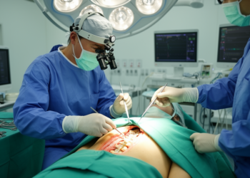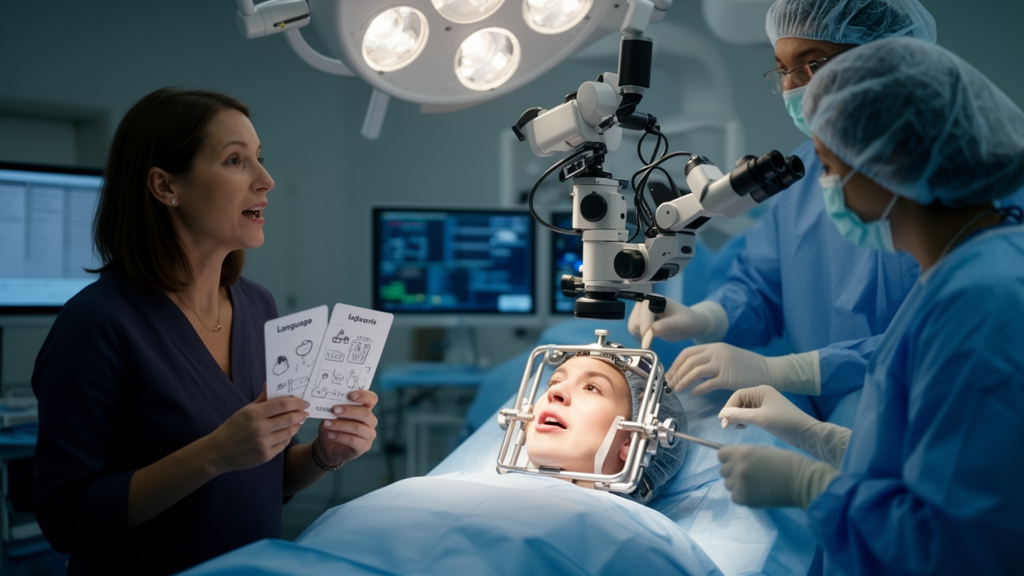
Neurosurgeons have long favored awake craniotomy for lesions sitting in or near the brain’s most precious real estate: the cortical and subcortical zones that control movement and language. By keeping patients sedated but rousable, and by testing their speech and motor function in real time while the surgeon works, the technique aims to remove as much lesion as possible without cutting through pathways the patient cannot afford to lose. A new retrospective study from a single Chinese center now offers a detailed, if cautiously framed, picture of what happens to such patients in the first days after surgery.
The study, published in the Chinese Neurosurgical Journal, followed 30 patients who underwent awake craniotomy for intracranial lesions involving motor or language functional cortical areas between January 2019 and November 2024 at the Second People’s Hospital of Changzhou. The authors, led by Yajie Song and Fang Liu, describe their work explicitly as a descriptive retrospective analysis, and they attach unusually strong caveats to their own findings, particularly the survival data. That restraint matters, because the cohort is small and pathologically mixed, and the outcome window they examined is brief.
The central result concerns the 48-to-72-hour postoperative window. Within that period, 30.0 percent of patients, nine of thirty, developed new or aggravated acute neurological dysfunction. The researchers deliberately split these changes into three categories: transient global decline, focal motor deficits, and isolated language impairment. The distinction is clinically important, because the two types carry very different prognostic weight.
Transient global decline dominated the picture. A drop of at least 10 points on the Karnofsky Performance Status scale occurred in 30.0 percent of patients, and a decline of at least 2 points on the Mini-Mental State Examination appeared in 26.7 percent. The authors attribute these early reductions largely to reversible perioperative factors: cerebral edema around the surgical site, systemic postoperative fatigue, and the lingering clearance of intraoperative sedatives such as remifentanil and dexmedetomidine. In other words, the patient who seems cognitively dulled two days after surgery is often not permanently injured but temporarily overwhelmed by the physiology of recovery.
Focal deficits, the surrogates most closely tied to irreversible injury, were far less common. New or worsened motor weakness occurred in 16.7 percent of patients, five of thirty, and de novo aphasia in only 10.0 percent, three of thirty. The authors read the low aphasia rate as evidence for the language-preserving capacity of intraoperative direct electrical stimulation mapping, a technique in which a bipolar probe delivers current at 2 milliamperes, titrated up to a maximum of 6, to identify cortical and subcortical functional boundaries. Resection is halted if consecutive positive stimulation signals are detected within 5 millimeters of the planned resection margin.
The mapping itself succeeded in most cases. Full intraoperative awake mapping was completed in 90.0 percent of patients, 27 of 30, while three patients required conversion to general anesthesia with incomplete functional mapping. The success rate, the authors note, aligns with published international multicenter cohorts. Interestingly, patients with successful mapping showed a numerically lower rate of acute neurological decline, 25.9 percent, compared with 66.7 percent among the small group in whom mapping failed. But with only three failures, the association fell short of statistical significance, with an odds ratio of 0.18, a 95 percent confidence interval of 0.01 to 2.34, and a P value of 0.20.
That null result extended across the board. Univariate logistic regression identified no statistically significant clinical predictors of acute postoperative neurological deterioration. Age of 55 years or older, preoperative Karnofsky score of 60 or above, preoperative Mini-Mental State Examination score of 27 or above, and high-grade glioma pathology all failed to correlate significantly with acute decline, with P values ranging from 0.24 to 0.72. The authors did not attempt multivariable modeling for the primary endpoint, citing the small sample size and the low event rate of severe focal deficits.
The cohort itself reflects the messy reality of neurosurgical practice rather than the filtered populations of landmark glioma trials. Of the 30 patients, whose mean age was 54.2 years with a standard deviation of 14.3, the pathology mix included 13 high-grade gliomas, eight glioblastomas and five anaplastic gliomas, seven low-grade gliomas, six cerebral metastases, three intracranial meningiomas, and one brain arteriovenous malformation. High-grade gliomas accounted for 43.3 percent of cases. Twenty-three patients were male and seven female. Before surgery, 76.7 percent had independent functional status with a Karnofsky score of at least 60, and 46.7 percent had normal baseline cognition with a Mini-Mental State Examination score of at least 27.
The survival analyses, which the authors repeatedly label exploratory and hypothesis-generating, produced no significant findings. Over follow-up completed by February 28, 2025, all-cause mortality reached 56.7 percent, 17 of 30 patients, with a median overall survival of 43.0 months and a 95 percent confidence interval of 30.5 to 55.5 months. Log-rank comparisons found no significant survival differences between high-grade and low-grade glioma, with median survival of 40.0 versus 45.0 months and a P value of 0.24, or between successful and failed mapping, 45.0 versus 35.0 months with a P value of 0.58. Univariate and multivariate Cox proportional hazards regression likewise identified no independent prognostic factors for all-cause mortality.
The limitations the authors enumerate are substantial and worth taking seriously. The pathological heterogeneity pools together diseases with fundamentally different biology and prognostic trajectories. Key covariates, including standardized volumetric extent of resection, precise lesion localization, and molecular glioma biomarkers such as IDH mutation status and 1p/19q codeletion, were unavailable in the retrospective records and could not enter the regression models. All neurological assessments were confined to the 48-to-72-hour window, so the study cannot distinguish transient perioperative dysfunction from sustained permanent injury, nor quantify how many patients with acute decline ultimately recovered. There was no matched general anesthesia control group, and excluding mapping-failure cases with incomplete endpoint data may have underestimated the true procedural risk of conversion. The authors conclude that large, prospective, pathology-stratified controlled trials with serial long-term cognitive, motor, and language assessments are needed before definitive claims about risk factors and long-term functional benefit can be made. Within those limits, the study’s practical message stands: early global dulling after awake craniotomy is common and usually reversible, while permanent language injury remains rare when direct electrical stimulation mapping is completed successfully.
Subject of Research: Short-term neurological outcomes and exploratory survival after awake craniotomy for intracranial lesions in eloquent brain areas
Article Title: Neurological outcomes and survival in patients with intracranial lesions undergoing awake craniotomy: a descriptive retrospective single-center study
Article References: Song, Y., Yang, K., Zhang, Y., Shen, L., Guo, Y., Yu, Z., & Liu, F. (2026). Neurological outcomes and survival in patients with intracranial lesions undergoing awake craniotomy: a descriptive retrospective single-center study. Chinese Neurosurgical Journal, 12(1), Article 26. https://doi.org/10.1186/s41016-026-00443-9
Image Credits: AI Generated
DOI: 10.1186/s41016-026-00443-9
Keywords: awake craniotomy, direct electrical stimulation mapping, eloquent cortex, glioma, aphasia, Karnofsky Performance Status, Mini-Mental State Examination, postoperative neurological deficits, overall survival, retrospective study, neurosurgery, cerebral edema
Cite Scienmag News
Ophelia Keating. (October 29, 2026). Awake craniotomy study finds transient decline common after eloquent-area surgery. Scienmag. https://scienmag.com/awake-craniotomy-study-finds-transient-decline-common-after-eloquent-area-surgery/
Ophelia Keating. "Awake craniotomy study finds transient decline common after eloquent-area surgery." Scienmag, 29 October 2026, https://scienmag.com/awake-craniotomy-study-finds-transient-decline-common-after-eloquent-area-surgery/. Accessed 29 October 2026.
Ophelia Keating. "Awake craniotomy study finds transient decline common after eloquent-area surgery." Scienmag. October 29, 2026. https://scienmag.com/awake-craniotomy-study-finds-transient-decline-common-after-eloquent-area-surgery/