In the rapidly evolving field of neuroimaging, Magnetic Resonance Imaging (MRI) has emerged as a foundational technology, enabling researchers to explore the intricate anatomy and functionality of the human brain. However, the utility of MRI data is intrinsically tied to its quality. The presence of noise or artifacts in raw MRI scans can severely compromise downstream analyses, potentially leading to false conclusions or masking genuine neural phenomena. Recognizing this challenge, a team of researchers has introduced a comprehensive and robust protocol for the quality control (QC) of unprocessed MRI data, leveraging an advanced tool known as MRIQC. This protocol promises to transform neuroimaging workflows by ensuring that only the highest fidelity images proceed to the preprocessing stages, thereby enhancing the reliability of scientific insights derived from MRI studies.
MRI data exists in several forms — anatomical (T1- and T2-weighted scans), functional (fMRI), and diffusion MRI — each providing distinct insights into brain structure or activity. The inherent complexity of these imaging modalities necessitates rigorous quality checks prior to any data preprocessing. Poor-quality scans can introduce variability into datasets, often in the form of noise or signal distortions, which may manifest either as spurious effects or as subtle obfuscations of true neurobiological signals. This methodological vulnerability underscores the urgent need for automated yet precise QC systems that can quickly flag substandard images according to predefined exclusion criteria. The described protocol not only addresses this need but also integrates seamlessly with typical high-performance computing environments common in contemporary research institutions.
At the heart of this approach is MRIQC, an open-source software suite designed to perform detailed visual and quantitative assessments of MRI datasets. MRIQC generates visual reports that portray both anatomical and functional scans through various quality metrics and artifacts detection systems. By systematically applying MRIQC across datasets, researchers can identify and exclude problematic scans before embarking on elaborate preprocessing pipelines. This step is critical, as preprocessing on poor-quality data can yield unreliable or irreproducible scientific results, thus compromising the integrity of entire research studies.
Installation and configuration of MRIQC, although straightforward, require a careful setup, especially when applied to large datasets. The researchers outline a stepwise installation procedure optimized for execution on high-performance computing (HPC) clusters, environments that facilitate parallel processing of hundreds or thousands of images. Configuring the dataset within MRIQC involves 30 to 45 minutes of active user engagement, ensuring that the software correctly interprets the imaging modalities and metadata. Subsequently, each scan undergoes automated quality metric computations, which require an average of 10 to 15 minutes of compute time per scan, depending on scanner parameters and image resolution. These timings highlight the balance between computational efficiency and thorough data scrutiny.
Following the automated generation of quality visualizations and metrics, the next crucial phase is the human-driven interpretation and annotation of these reports. This hybrid QC method, combining algorithmic rigor with expert visual verification, helps to capture subtle imperfections that might escape purely automated systems. To streamline this annotation process, the protocol introduces the ‘rating widget,’ a specialized interface tool designed for rapid, accurate labeling of image quality. This widget minimizes the risk of bookkeeping errors, a common pain point in large-scale neuroimaging studies, and accelerates the decision-making workflow to between one and five minutes per participant — a significant time-saving compared to conventional methods.
A particularly compelling strength of this protocol lies in its preventative impact on data acquisition quality. Early detection of scanning faults allows imaging teams to address and rectify hardware or protocol-related issues in real time, rather than discovering these problems post-hoc during data analysis. This proactive feedback loop is invaluable for large-scale studies or longitudinal research where repeated scans over extended periods are common, and the retrospective exclusion of flawed data could severely erode statistical power or introduce bias.
Moreover, the emphasis on unprocessed data quality assessment aligns with growing awareness in the neuroscience community regarding reproducibility and data transparency. Suboptimal datasets, if processed without appropriate quality filters, contribute to the “noise floor” within brain imaging literature, complicating meta-analyses and hindering the development of robust neurobiological models. Tools like MRIQC democratize access to standardized QC procedures, enabling research consortia to harmonize quality thresholds and share best practices across diverse laboratories and scanners.
While the protocol was originally optimized for research-grade scanners generating high-resolution neuroimaging data, the underlying principles can be adapted for clinical or developmental imaging studies, where rapid yet precise quality assessment is equally vital. The scalability of MRIQC on HPC infrastructures ensures that large public datasets, encompassing thousands of subjects, can be processed efficiently without sacrificing individual scan scrutiny. As neuroimaging datasets continue to expand exponentially, such scalable QC pipelines become indispensable.
Beyond artifact detection and noise quantification, MRIQC’s metrics provide insights into underlying scanner performance and protocol adherence. Common MRI artifacts such as signal dropouts, ghosting, or geometric distortions manifest clearly in visual reports, allowing teams to trace their origins back to scanner malfunctions or participant-related factors like head movement. This diagnostic capability enhances longitudinal data reliability and fosters methodological improvements in image acquisition protocols.
Importantly, the protocol situates quality control as an integral initial step rather than an optional add-on or post hoc correction. This mindset shift encourages a culture of quality consciousness from data inception through to publication. Such rigorous QC protocols can mitigate the risk of spurious findings, amplifying the robustness of neuroimaging as a tool for unraveling brain-behavior relationships and neurological disorders.
Industry and academic collaborations stand to benefit immensely from this advancement. The researchers’ detailed procedural guidance, enriched with precise timing estimates and implementation notes, lowers the entry barrier for laboratories new to MRIQC or large-scale MRI datasets. By fostering widespread adoption of these best practices, the protocol promotes a standard benchmark for MRI data quality, facilitating cross-study comparisons and integrative analyses.
Ultimately, the approach described by Hagen et al. exemplifies how computational power, combined with nuanced expert evaluation, can elevate data integrity in complex biomedical imaging. As brain research accelerates into the era of big data and machine learning, maintaining stringent quality assurance will be pivotal for unlocking the full potential of MRI datasets and translating neuroimaging discoveries into clinical and societal impact.
The fusion of algorithmic automation and human insight embodied in this protocol paves the way for more reproducible, transparent, and credible neuroscience research. Ensuring that substandard MRI scans are identified and excluded at the earliest stages preserves the fidelity of downstream analyses and prevents errant conclusions that could misdirect future investigations. This methodology marks a critical step forward in the ongoing quest to decode the mysteries of the human brain with precision and confidence.
Researchers and clinicians interested in implementing robust MRI quality control can access MRIQC and its documentation via the project’s web platform, enabling a community-driven evolution of QC standards tailored to emerging technologies and research needs. The protocol’s integration with established high-performance cluster computing environments also future-proofs quality control pipelines against the growing scale and ambition of neuroimaging endeavors.
In summary, as neuroimaging expands its frontiers, the establishment of rigorous, efficient, and reproducible quality control protocols will underpin credible scientific progress. The novel approach presented here embodies this imperative, positioning MRIQC as a vital resource in the ongoing effort to optimize MRI data fidelity, enhance analytical accuracy, and deepen our understanding of the brain’s complex architecture and function.
Subject of Research: Quality assessment and control of unprocessed anatomical, functional, and diffusion MRI of the human brain
Article Title: Quality assessment and control of unprocessed anatomical, functional and diffusion MRI of the human brain using MRIQC
Article References:
Hagen, M.P., Provins, C., MacNicol, E. et al. Quality assessment and control of unprocessed anatomical, functional and diffusion MRI of the human brain using MRIQC. Nat Protoc (2026). https://doi.org/10.1038/s41596-026-01352-y
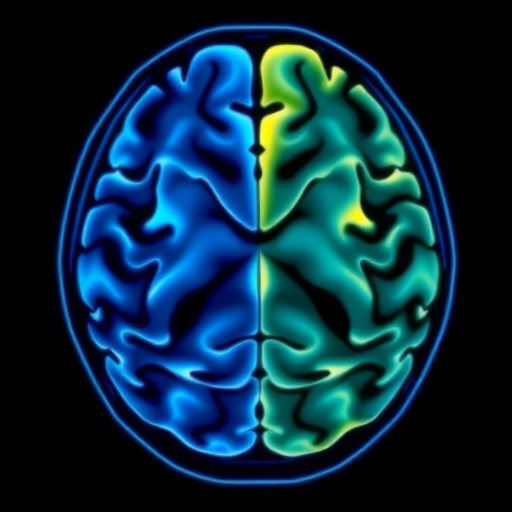
Image Credits: AI Generated
