


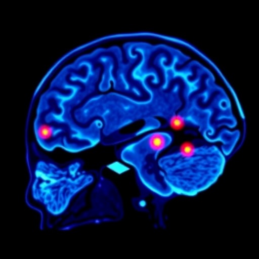
A recent meta-analysis focusing on Alzheimer’s disease biomarkers has introduced refined insights into the interpretation of amyloid PET imaging. By pooling individual participant data from multiple studies, researchers honed in on Centiloid thresholds—a quantitative measure used to standardize amyloid PET scans across different imaging centers and tracers. Their results delineate two key positivity cutoffs: approximately 18 Centiloids for data-driven determinations and around 27 Centiloids when relying on visual reads by experts.
This convergence of cutoffs offers a more unified framework for identifying amyloid positivity, a crucial factor in diagnosing and monitoring Alzheimer’s pathology. Importantly, the study highlights a “gray zone” spanning 11 to 26 Centiloids. Scans falling within this intermediate range demand caution, indicating that diagnostic interpretation should be context-dependent and consider additional clinical and biomarker information.
The findings stem from a rigorous double-cutoff analysis designed to parse out sensitivity and specificity trade-offs inherent in amyloid PET scanning. This dual approach allows for a more nuanced understanding of how different thresholds influence the classification of amyloid burden, addressing longstanding debates in neuroimaging standardization.
Amyloid PET imaging is a pivotal tool for detecting beta-amyloid plaques in vivo, enabling earlier and more accurate Alzheimer’s diagnoses prior to symptom onset. However, varying imaging protocols and interpretative criteria have historically challenged consistency across studies and clinical centers. The meta-analytic approach in this study circumvents these limitations by leveraging harmonized participant-level data, yielding robust cutoffs that can be broadly applied.
Moreover, the implication that scans with Centiloid values in the lower intermediate range should be interpreted cautiously could impact both clinical practice and research, particularly in trials targeting preclinical or prodromal Alzheimer’s stages. It suggests that reliance solely on amyloid PET positivity may risk misclassification without complementary assessments such as cognitive testing or cerebrospinal fluid biomarkers.
Published in JAMA to coincide with the Alzheimer’s Association International Conference, this work signifies a critical step toward standardizing amyloid PET quantification. The research team, led by Dr. Ganna Blazhenets, underscores the importance of precision in neurodegenerative disease imaging, recommending that interpretation protocols incorporate these refined cutoffs to enhance diagnostic accuracy.
As quantitative imaging advances, integration of standardized Centiloid thresholds will facilitate harmonized data sharing and meta-analyses in the Alzheimer’s research community. It also paves the way for developing tailored interventions by reliably stratifying patient amyloid status.
In summary, this meta-analytic study refines amyloid PET positivity thresholds to 18 Centiloids for data-driven assessment and 27 Centiloids for visual interpretation, with a cautious zone between 11 and 26 Centiloids. These findings emphasize nuanced diagnostic scrutiny in scans within this gray area and promise to advance clinical and research practices in Alzheimer’s disease.
Cite Scienmag News
Ophelia Keating. (July 13, 2026). Amyloid PET Quantification Enhances Alzheimer’s Disease Diagnosis Accuracy. Scienmag. https://scienmag.com/amyloid-pet-quantification-enhances-alzheimers-disease-diagnosis-accuracy/
Ophelia Keating. "Amyloid PET Quantification Enhances Alzheimer’s Disease Diagnosis Accuracy." Scienmag, 13 July 2026, https://scienmag.com/amyloid-pet-quantification-enhances-alzheimers-disease-diagnosis-accuracy/. Accessed 4 September 2026.
Ophelia Keating. "Amyloid PET Quantification Enhances Alzheimer’s Disease Diagnosis Accuracy." Scienmag. July 13, 2026. https://scienmag.com/amyloid-pet-quantification-enhances-alzheimers-disease-diagnosis-accuracy/


