


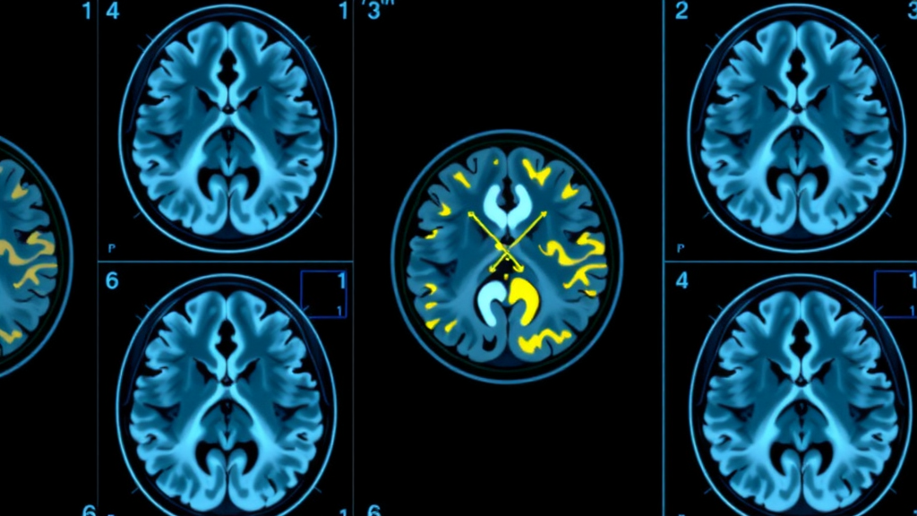
For decades, brain imaging specialists have treated the cerebellum as the silent, trustworthy backdrop against which Alzheimer’s disease can be measured. When researchers quantify amyloid-beta plaques using positron emission tomography, they routinely divide the tracer signal in the cerebral cortex by the signal in a reference region assumed to be free of amyloid pathology. The whole cerebellum has been the standard choice, enshrined in the Centiloid scale that harmonizes amyloid PET measurements across tracers, centers, and clinical trials. A new study now shows that this foundational assumption fails in a clinically meaningful subset of patients, with consequences that ripple through diagnosis, trial monitoring, and the assessment of anti-amyloid therapies.
The research, led by Meixi Wang, Sheng Bi, and colleagues at Xuanwu Hospital of Capital Medical University in Beijing and published in the European Journal of Nuclear Medicine and Molecular Imaging, examined 359 amyloid-positive individuals imaged with the widely used tracer 18F-Florbetapir. Among them, 52 showed visible amyloid-beta deposition in the cerebellar cortex itself, while 307 did not. The team combined PET imaging, structural MRI, and cerebrospinal fluid biomarker analysis to ask a deceptively simple question: what happens to standard amyloid measurements when the reference region is itself contaminated with amyloid?
The answer was striking. Participants with cerebellar cortical amyloid deposition had significantly lower Centiloid values than those without, despite all being amyloid-positive. The mechanism involves a dual bias: tracer uptake in the whole cerebellum was elevated by direct amyloid binding in the cerebellar cortex, while cortical tracer uptake was paradoxically reduced, possibly reflecting advanced-stage processes such as synaptic loss and declining soluble amyloid in cerebrospinal fluid. Dividing a lower cortical signal by a higher cerebellar signal systematically underestimates the true cerebral amyloid burden, potentially misleading clinicians about how much plaque a patient actually carries.
The quantitative details matter. Whole-cerebellum standardized uptake values were significantly higher in the cerebellar-positive group, cortical uptake values were significantly lower, and Centiloid values dropped dramatically, with bootstrap analysis confirming the differences were far from chance. By contrast, uptake in the pons did not differ significantly between groups, and when cerebral white matter was used as the denominator, the resulting SUVR showed no significant between-group difference at all. White matter, long considered a secondary option, emerged as the only reference region that remained stable in the face of cerebellar pathology.
TheCentiloid scale, anchored at zero for young healthy controls and 100 for typical Alzheimer’s patients, has become the lingua franca of amyloid quantification. It underpins threshold decisions in clinical practice and serves as a primary endpoint in trials of anti-amyloid antibodies such as lecanemab, donanemab, and aducanumab. The new findings suggest that in patients with cerebellar involvement, Centiloid values cannot be taken at face value. A patient whose Centiloid score appears to fall below the conventional amyloid-positivity threshold of 30 may still carry substantial cortical plaque burden, simply because the denominator of the ratio has been inflated by cerebellar amyloid.
To help clinicians detect this problem, the team established and validated a quantitative diagnostic threshold. Using cerebellar SUVR normalized to white matter, receiver operating characteristic analysis yielded an optimal cutoff of 0.428, with an area under the curve of 0.931, sensitivity of 86.5 percent, and specificity of 86.3 percent. In an internal validation subset of 30 patients with equivocal visual readings, agreement between the quantitative threshold and expert visual assessment was substantial, with a Cohen’s kappa of 0.856. The negative predictive value of 97.4 percent was particularly strong, positioning the metric as an effective screening tool for ruling out cerebellar amyloid involvement.
The longitudinal component of the study adds a provocative therapeutic dimension. Two amyloid-positive patients with cerebellar cortical deposition were treated with lecanemab and followed with repeat PET at seven and eleven months. In both, Centiloid values decreased substantially over time, yet cerebellar SUVR normalized to white matter rose, remaining above the 0.428 threshold throughout follow-up. In one patient, the Centiloid value fell from 70.9 to 22.4, crossing below the recommended positivity threshold even though visual assessment confirmed persistent cortical amyloid and unchanged cerebellar deposition. Reading such scans through a Centiloid lens alone could falsely suggest near-complete amyloid clearance.
The persistence of cerebellar amyloid despite antibody therapy has a plausible biological explanation. Unlike the cerebral cortex, where compact cored plaques predominate, the cerebellum is characterized by diffuse amyloid deposits. Autopsy findings from the historic AN-1792 immunization trial showed that cerebellar diffuse plaques resisted clearance even when neocortical plaques were removed. Because passive antibodies such as lecanemab engage microglial clearance pathways that depend on Fc-gamma receptor-mediated phagocytosis, pathways more effective against compact plaques than diffuse ones, the cerebellar signal may simply not respond to treatment in the same way as the cortex. The new data are consistent with that framework.
Cerebellar amyloid is not a rarity confined to exotic cases. According to the Thal staging system, cerebellar cortical involvement marks phase five of Alzheimer’s pathology, the most advanced stage. In autosomal dominant Alzheimer’s disease caused by Presenilin-1 mutations, PET studies have detected cerebellar amyloid roughly a decade before symptom onset. And a recent autopsy study in the oldest-old suggested cerebellar cortical deposition may be more common in aged clinical populations than previously appreciated, precisely the group now being considered for anti-amyloid therapy. As treatment eligibility increasingly hinges on quantitative amyloid thresholds, accurate measurement in these patients becomes a pressing clinical concern.
The authors caution that the 0.428 threshold was derived in a single-center cohort, the longitudinal analysis included only two patients, and the findings require replication with other amyloid tracers and in independent multicenter samples. Nevertheless, the practical implications are immediate. Where cerebellar amyloid is suspected, white matter offers a robust alternative reference region, and the cerebellar SUVR threshold provides a standardized, readily implementable check on cerebellar status. The study also redefines what makes a reference region valid: freedom from amyloid cannot be assumed globally, but must be verified for each patient and disease stage. As quantitative amyloid PET becomes central to the era of disease-modifying Alzheimer’s therapy, ensuring that the yardstick itself is clean may prove just as important as the drug being measured.
Subject of Research: Bias in 18F-Florbetapir amyloid PET quantification caused by cerebellar cortical amyloid-beta deposition
Article Title: Cerebellar cortical amyloid deposition biases 18F-Florbetapir PET quantification with whole cerebellum as reference region
Article References: Wang, M., Bi, S., Xue, H., Guan, L., Wang, Y., Zhang, X., Liu, X., Xu, X., Zhang, C., Qi, Z., Zhao, Z., Yan, S., & Lu, J. (2026). Cerebellar cortical amyloid deposition biases 18F-Florbetapir PET quantification with whole cerebellum as reference region. European Journal of Nuclear Medicine and Molecular Imaging. https://doi.org/10.1007/s00259-026-08140-6
Image Credits: AI Generated
DOI: 10.1007/s00259-026-08140-6
Keywords: Alzheimer's disease, amyloid-beta PET, 18F-Florbetapir, cerebellar amyloid, Centiloid scale, reference region, white matter, SUVR, lecanemab, quantification bias, nuclear medicine, diagnostic threshold
Cite Scienmag News
Ophelia Keating. (September 23, 2026). Amyloid in the Cerebellum Is Quietly Corrupting Alzheimer’s PET Scans. Scienmag. https://scienmag.com/amyloid-in-the-cerebellum-is-quietly-corrupting-alzheimers-pet-scans/
Ophelia Keating. "Amyloid in the Cerebellum Is Quietly Corrupting Alzheimer’s PET Scans." Scienmag, 23 September 2026, https://scienmag.com/amyloid-in-the-cerebellum-is-quietly-corrupting-alzheimers-pet-scans/. Accessed 5 October 2026.
Ophelia Keating. "Amyloid in the Cerebellum Is Quietly Corrupting Alzheimer’s PET Scans." Scienmag. September 23, 2026. https://scienmag.com/amyloid-in-the-cerebellum-is-quietly-corrupting-alzheimers-pet-scans/


