


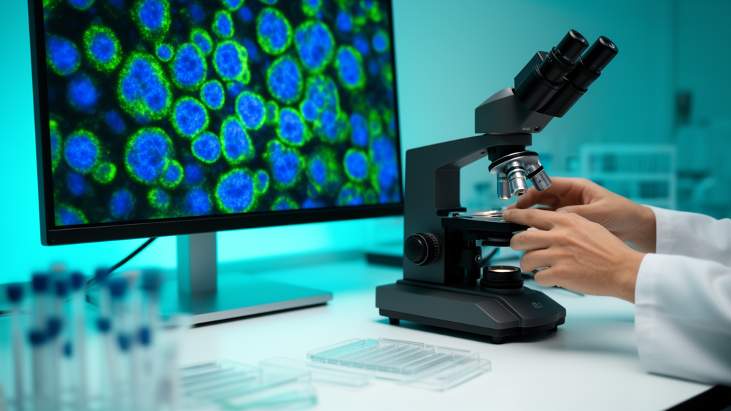
Fluorescence microscopy has long been one of the most powerful windows into the living cell, allowing biologists to tag specific molecules with glowing labels and watch them move, interact and organize across space and time. Yet the technique has always been constrained by a stubborn set of trade-offs. Pushing for higher resolution often means blasting delicate specimens with more light and slowing acquisition to a crawl. Chasing faster dynamics sacrifices signal quality, and imaging larger volumes multiplies both problems at once. A new review published in PhotoniX Life argues that deep learning is now fundamentally rewriting those trade-offs, offering researchers a way to extract more information from fewer, noisier and faster-acquired images than ever before.
The review, authored by researchers affiliated with the Chinese Society for Optical Engineering, systematically maps the applications of artificial intelligence across four major directions: imaging quality, imaging speed, imaging throughput and intelligent analysis. Rather than treating neural networks as a single monolithic tool, the authors begin by dissecting the mainstream architectures that underpin modern computational microscopy, including convolutional neural networks, U-Net, generative adversarial networks, Transformers and diffusion models. Each architecture carries distinct strengths. Convolutional networks excel at extracting local spatial features from pixel grids, making them natural fits for denoising and segmentation. Generative adversarial networks can synthesize plausible high-quality outputs from degraded inputs, while Transformers capture long-range dependencies across an image. Diffusion models, the newest entrants, generate reconstructions through iterative denoising steps and have shown remarkable fidelity in restoration tasks. The review compares these approaches in terms of feature extraction capability, computational cost and suitability for specific microscopy problems, giving experimentalists a practical framework for choosing the right tool.
The first application domain, imaging quality, is where deep learning has made its deepest inroads. Super-resolution reconstruction, the computational expansion of images beyond the diffraction limit of the optical system, has been transformed by neural networks trained to infer fine structural details that conventional optics blur away. Image restoration pipelines can rescue low-signal frames that would once have been discarded, effectively letting microscopes operate at lower illumination levels and reducing phototoxicity in living specimens. Digital aberration correction adds another layer of sophistication, with algorithms compensating for optical imperfections that vary across the field of view. Perhaps the most important trend the review identifies here is a decisive move away from complete dependence on paired training data, in which every low-quality image must be matched to a corresponding high-quality ground truth. Self-supervised and zero-shot approaches now exploit statistical information already contained within experimental data, while physics-informed methods embed explicit knowledge of the imaging process directly into model training, anchoring reconstructions to the actual physics of light propagation rather than to statistical guesswork.
Imaging speed represents the second pillar. Many biological processes of interest, from calcium signaling in neurons to the rapid remodeling of cytoskeletal filaments, unfold on timescales that conventional acquisition strategies struggle to capture. Neural networks address this bottleneck by reducing the number of raw measurements required for a faithful reconstruction, accelerating localization algorithms that pinpoint single fluorescent molecules, and in some cases replacing repeated mechanical scanning or filter switching entirely with computational prediction. Instead of physically moving a stage or rotating a filter wheel through multiple acquisition cycles, the microscope captures a reduced set of observations and the network fills in the rest. For rapidly changing biological dynamics, this shift from mechanical to computational acquisition can mean the difference between observing a transient molecular event and missing it altogether.
The third direction, imaging throughput, tackles the challenge of scale. Modern biology increasingly demands volumetric imaging of entire organoids, cleared tissue sections or whole organisms, tasks that generate terabytes of data and strain both optical hardware and computational pipelines. The review describes how deep learning has been combined with light-field imaging, which captures three-dimensional information in a single snapshot, and with field-dependent aberration correction, which maintains image quality across wide volumes. Parallel data processing strategies accelerated by neural networks then expand the effective imaging volume and speed up the reconstruction of large-scale three-dimensional datasets. Together these approaches point toward microscopes that can survey biological systems at scales previously impractical, without proportionally increasing acquisition time or light exposure.
Beyond the reconstruction of images themselves, the fourth domain concerns biological interpretation, and it is here that the review raises some of its most consequential cautions. Deep learning now enables automated cell and subcellular segmentation, converting raw micrographs into quantitative measurements of morphology, count and spatial organization. Even more strikingly, virtual fluorescent labeling techniques can generate computationally predicted fluorescence from label-free inputs, inferring where specific molecular structures would appear without any staining at all. These capabilities dramatically reduce labeling demands and spare specimens from the perturbations that fluorescent tags can introduce. But the review is unambiguous on a critical point: computationally predicted signals are not equivalent to experimentally measured molecular signals. A network that convincingly paints mitochondria onto an unlabeled image is producing an informed inference, not a direct observation, and treating the two as interchangeable risks contaminating downstream biological conclusions.
This concern feeds into the central message of the entire review, namely that visual improvement alone is an insufficient standard for AI-assisted microscopy. An image that looks crisp and convincing to the human eye may nonetheless contain hallucinated structures, smoothed-over details or artifacts that a network invented to satisfy its training objectives. Reliable AI-assisted microscopy, the authors argue, must preserve genuine biological structure, generalize across different samples and instruments, and keep pace with experimental acquisition speeds. A model that performs beautifully on one microscope’s data but fails on another’s is of limited scientific value, and the review highlights limited cross-platform generalization as one of the field’s most pressing unsolved problems, alongside the strong dependence of current methods on training data quality and quantity.
Evaluation practice, the review contends, must evolve accordingly. Standard image-quality metrics such as peak signal-to-noise ratio and structural similarity index, while convenient, capture only coarse statistical agreement between a reconstruction and its reference. They say little about whether fine subcellular features have been faithfully recovered or subtly fabricated. The authors call for evaluation frameworks that go beyond these familiar metrics to include spatial resolution measurements, local reconstruction fidelity checks, and performance on downstream biological tasks such as segmentation or tracking, which test whether a reconstruction actually supports the scientific question being asked. Wherever feasible, they add, computational results should be validated against independent experimental measurements, closing the loop between algorithmic output and biological reality.
Looking toward the future, the review identifies three key directions for the field. The first is the deeper integration of physical imaging models with data-driven learning, so that neural networks are constrained by the known physics of fluorescence excitation, emission and detection rather than learning everything from scratch. The second is the co-design of optical hardware and algorithms, building microscopes and networks together so that each is optimized for the other, rather than bolting software onto instruments designed for purely analog workflows. The third is the establishment of standardized evaluation protocols, giving the community shared benchmarks against which competing methods can be fairly compared and their limitations honestly assessed.
The trajectory the review sketches is one in which deep learning graduates from a post-processing afterthought to an integral component of the microscope itself, embedded in the acquisition pipeline from the moment photons strike the detector. For experimental biologists, the practical guidance is clear: understand what the algorithms can and cannot do, choose methods appropriate to the imaging task at hand, and evaluate reconstructions with the same rigor applied to any other experimental measurement. As intelligent fluorescence microscopy matures, the laboratories that benefit most will be those that pair enthusiasm for artificial intelligence with a sober respect for its limits, ensuring that the images of tomorrow are not only beautiful but biologically true.
Subject of Research: Applications of deep learning in fluorescence microscopic imaging
Article Title: Deep learning is reshaping fluorescence microscopy from image acquisition to biological analysis
Article References: Deep learning is reshaping fluorescence microscopy from image acquisition to biological analysis. (n.d.). Original publication
Image Credits: AI Generated
DOI: Not provided
Keywords: deep learning, fluorescence microscopy, super-resolution, image denoising, neural networks, cell segmentation, virtual labeling, light-field imaging, aberration correction, 3D imaging, physics-informed models, PhotoniX Life
Cite Scienmag News
Blake Davidson. (October 15, 2026). AI transforms fluorescence microscopy from image capture to biological discovery. Scienmag. https://scienmag.com/ai-transforms-fluorescence-microscopy-from-image-capture-to-biological-discovery/
Blake Davidson. "AI transforms fluorescence microscopy from image capture to biological discovery." Scienmag, 15 October 2026, https://scienmag.com/ai-transforms-fluorescence-microscopy-from-image-capture-to-biological-discovery/. Accessed 15 October 2026.
Blake Davidson. "AI transforms fluorescence microscopy from image capture to biological discovery." Scienmag. October 15, 2026. https://scienmag.com/ai-transforms-fluorescence-microscopy-from-image-capture-to-biological-discovery/


