
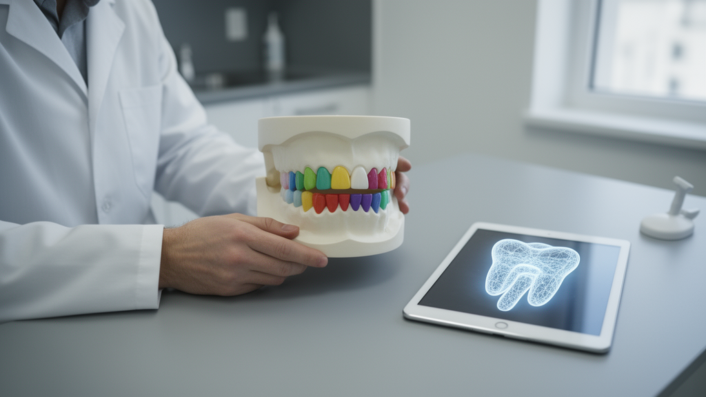
Artificial intelligence has quietly transformed one of dentistry’s most tedious chores: tracing the outlines of every tooth in a medical image. A new systematic review published in Discover Imaging pulls together a decade of research on automatic tooth segmentation, cataloguing 72 representative studies published between 2016 and 2025 and twelve open-source datasets spanning panoramic radiographs, cone-beam computed tomography, intraoral scans and cephalometric radiographs.
The stakes are more practical than they might sound. Accurate segmentation of teeth is a prerequisite for disease diagnosis, treatment planning and the broader digitalisation of dental care. Before deep learning, clinicians relied on manual or semi-automatic tracing, which was time-consuming and inconsistent. Traditional mathematical approaches such as thresholding, region growing, watershed algorithms, Snake curves and level-set methods still appear in the literature, and the review notes they retain value as preprocessing steps and in small-sample settings, but their accuracy typically ranges between 70 and 80 percent, short of clinical precision requirements.
The authors searched PubMed, IEEE Xplore and Web of Science for articles containing the keywords “teeth segmentation” or “tooth segmentation,” collecting 2,472 records from 1997 to 2025. After removing 1,499 duplicates with automated tools, 973 articles remained for bibliometric analysis in CiteSpace, a Java-based tool for visualising research trends. Screening then excluded non-English publications, non-peer-reviewed work, studies without standard evaluation metrics, and those using extremely small training samples without public datasets. The final 72 studies split into 14 on fundamental network architectures, 23 on two-dimensional segmentation and 35 on three-dimensional segmentation, cross-checked independently by two authors.
The bibliometric analysis traces a striking shift in vocabulary. The keyword co-occurrence map generated 559 nodes and 1,967 links, with deep learning, artificial intelligence, segmentation and classification emerging as dominant hotspots. Before 2010, terms like variational level set, CT and active contours showed significant bursts before declining. Between 2010 and 2019, CBCT and automatic segmentation grew explosively. Since 2020, deep learning, dental panoramic radiographs, instance segmentation and semantic segmentation have prevailed, mirroring the field’s migration from hand-crafted mathematics to learned representations.
The review distinguishes two fundamental task types. Semantic segmentation, sometimes called binary segmentation, simply separates tooth from non-tooth regions, but it struggles when teeth touch or interdental spaces are narrow, because it cannot delineate individual teeth. Instance segmentation assigns a distinct label to each tooth, typically through multi-stage architectures combining detection and segmentation, and delivers the precise per-tooth boundaries that clinical applications demand. A third, multi-class approach labels each tooth according to the Federation Dentaire Internationale numbering standard, though the authors note this convention has yet to achieve widespread adoption.
Data availability is a major constraint on the field. The team identified 56 open-source dental imaging datasets on platforms such as Kaggle and Crowdflower and included twelve after screening: six panoramic radiograph collections, four CBCT collections, one intraoral scan dataset and one cephalometric radiograph dataset. Medical images are inherently private, and annotation is expensive; each CBCT slice requires manual tooth marking by trained specialists, and quality depends heavily on annotator expertise. Among the highlighted resources are CTooth and its extension CTooth+, the first large-scale open 3D dental CBCT datasets for voxel segmentation, the Multi-center CBCT Dataset with 150 annotated scans drawn from 4,215 patients across multiple institutions, the ToothFairy 2 dataset with 42 annotation categories used in a MICCAI 2024 challenge, and the Teeth3DS intraoral scan dataset covering 900 patients with annotations of 23,999 teeth.
On the algorithmic side, the review organises the field by backbone architecture. U-Net’s encoder-decoder design with skip connections remains the workhorse, excelling when training data are limited but capturing global context poorly. Mask R-CNN couples object detection with mask prediction for robust instance segmentation, though its computational demands confine it mostly to 2D tasks. Vision Transformers model long-range dependencies through self-attention but carry quadratic computational complexity, prompting hybrid designs such as TransUNet and nnFormer that blend convolutional local detail with Transformer global context. The newest entrant is Mamba, a state space model achieving linear complexity through a selective scan mechanism; derivatives like U-Mamba and VMamba are already appearing in medical segmentation, and the T-Mamba variant, which integrates frequency-domain features, reached a Dice coefficient of 93.60 percent on the public Multi-center CBCT Dataset.
The reported performance numbers illustrate steady progress, though the review is careful to frame them within their specific test conditions. On panoramic radiographs, a two-stage network combining YOLOv8 localisation with a bounding-box-guided U-Net improved average Dice scores by roughly 10 to 15 percent over conventional U-Net, particularly for multi-rooted molars and premolars. A federated learning study across nine international centres, using 4,177 private panoramic images, achieved an average sensitivity of 85.20 percent for instance segmentation while keeping raw images stored locally, a result the authors highlight as a path around data-privacy barriers. In three dimensions, two-stage pipelines that first localise tooth regions and then refine boundaries dominate, with reported Dice coefficients frequently exceeding 94 percent, including 96.2 percent for a hierarchical CNN on a 20-patient CBCT set and 94.1 percent for a V-Net-based hierarchy evaluated on the Multi-center CBCT Dataset.
The review also confronts the field’s stubborn technical obstacles. Metal crowns and implants produce streaking artefacts in CBCT images that distort grey values, causing tissue misclassification near metal objects and degrading segmentation accuracy. Intraoral scanners capture external crown and gingival surfaces with high precision but cannot see inside teeth and may accumulate stitching errors across full-arch scans, which is why some teams now fuse CBCT and IOS data to combine internal bone structure with accurate external surfaces. Evaluation practice itself is fragmented: overlap-based metrics such as Dice and IoU, distance-based measures in millimetres like Hausdorff distance, and volume-based metrics in cubic millimetres are all in use, with Dice preferred because teeth occupy a small fraction of each image and Dice is sensitive to small target regions.
The authors are candid about their own limitations. Because of heterogeneity among the included studies and the lack of publicly available data, no formal risk-of-bias assessment or certainty-of-evidence evaluation was performed; a narrative synthesis was used instead. They also note that varying datasets, annotation standards and the absence of external validation restrict comparability across studies, and that dataset standardisation efforts, such as the nnU-Net framework’s data conventions, remain little adopted in dental imaging. Their conclusions point toward standardised acquisition protocols, better generalisation across imaging devices with differing window settings, automated multimodal fusion rather than manual registration, and continued growth of federated learning for multi-institutional collaboration. On that trajectory, they argue, automatic dental segmentation is gradually positioning itself as a vital component of digital dentistry.
Subject of Research: A systematic review of deep learning methods for automatic tooth segmentation on dental medical imaging data.
Article Title: A review of tooth AI segmentation on medical data
Article References: Zhang, H., Yuan, T., Li, T., Du, J., Ye, M., & Jiang, Q. (2025). A review of tooth AI segmentation on medical data. Discover Imaging, 2(1), Article 17. https://doi.org/10.1007/s44352-025-00021-2
Image Credits: AI Generated
DOI: 10.1007/s44352-025-00021-2
Keywords: tooth segmentation, deep learning, dental imaging, CBCT, panoramic radiographs, U-Net, Mask R-CNN, Vision Transformer, Mamba, open-source datasets, digital dentistry, systematic review
Cite Scienmag News
Blake Davidson. (October 29, 2026). AI Tooth Segmentation Review Maps a Decade of Deep Learning Progress. Scienmag. https://scienmag.com/ai-tooth-segmentation-review-maps-a-decade-of-deep-learning-progress/
Blake Davidson. "AI Tooth Segmentation Review Maps a Decade of Deep Learning Progress." Scienmag, 29 October 2026, https://scienmag.com/ai-tooth-segmentation-review-maps-a-decade-of-deep-learning-progress/. Accessed 30 October 2026.
Blake Davidson. "AI Tooth Segmentation Review Maps a Decade of Deep Learning Progress." Scienmag. October 29, 2026. https://scienmag.com/ai-tooth-segmentation-review-maps-a-decade-of-deep-learning-progress/