


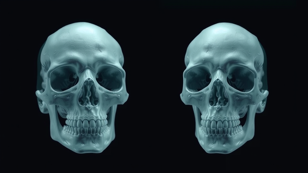
For decades, forensic artists have painstakingly sculpted clay over skulls, using population-averaged measurements of soft-tissue thickness to conjure a likeness of the dead. The results are often haunting, sometimes effective, and always tinged with subjectivity. Now, researchers in Vietnam have shown that a deep neural network can perform this delicate task automatically, learning to drape virtual flesh onto bone in a fully three-dimensional pipeline that could reshape how investigators generate leads in unidentified-body cases.
In a study published in the International Journal of Legal Medicine, Nguyen Tuan Kiet and Nguyen Truong Thinh of the University of Economics Ho Chi Minh City describe a skull-conditioned 3D U-Net that infers the facial soft-tissue envelope directly from cranial geometry. The work is deliberately framed as a feasibility-focused concept study rather than a finished forensic instrument, but its central result is striking: on an independent test set, the model achieved a mean Dice coefficient of 0.93, with a 95th-percentile Hausdorff distance of just 2.8 millimeters, indicating that the reconstructed faces closely hugged the true tissue boundaries in most regions.
The problem the researchers tackle is famously ill-posed. A skull does not uniquely determine the face that once covered it. Bone anchors certain regions firmly—forehead, nasal bridge, and the rims of the eye sockets are dictated largely by the geometry beneath—while the cheeks, jawline, and neck are dominated by fat, muscle, and connective tissue whose thickness varies enormously between individuals. Mathematically, this means the mapping from skull to face is one-to-many: a single cranium could sit beneath many anatomically plausible faces. Traditional manual reconstruction grapples with this ambiguity using tissue-depth pegs derived from population averages, a method that is reproducible only in part and inevitably biased toward the statistical middle.
The new framework reframes facial reconstruction as a supervised volumetric inference task. Each training example consists of paired three-dimensional volumes on an isotropic voxel grid: a binary skull mask as input and the corresponding outer facial soft-tissue envelope as output. The network learns a voxel-to-voxel mapping between them, producing a probability map in which every voxel carries a score reflecting the likelihood that it belongs to facial tissue. Crucially, the model receives no demographic hints whatsoever—no age, sex, ancestry, or body-mass index—so everything it infers must come from the shape of the bone itself. The authors argue this design choice is particularly appropriate for forensics, where such auxiliary information is frequently unavailable or unreliable.
Building the dataset demanded careful curation. The team collected 38 paired skull-and-face computed tomography volumes from clinical and forensic repositories, excluding cases with severe pathological deformity, acute trauma, or invasive implants that would confound the natural relationship between bone and soft tissue. Using Hounsfield Unit thresholds, they segmented bone at 250 HU or above and soft tissue in a range from minus 200 to 300 HU, then applied morphological closing and hole filling to guarantee topologically clean masks. All volumes were aligned to the Frankfort Horizontal Plane, resampled to uniform isotropic resolution, and resized to fixed grids of 192 by 192 by 192 voxels. With only 30 training cases and four each for validation and testing, the scale is modest, and the authors are candid that population-level generalization remains unproven.
Architecturally, the team resisted the temptation to innovate. They deliberately deployed a vanilla 3D U-Net—a symmetric encoder-decoder with four resolution levels, 3-by-3-by-3 convolutional kernels, LeakyReLU activations, and skip connections that shuttle fine geometric detail from encoder to decoder. The point, they explain, is to establish a principled baseline in which any observed performance can be attributed to the volumetric problem formulation rather than to architectural engineering. The output layer reduces the feature maps to a single probability channel through a 1-by-1-by-1 convolution and sigmoid activation.
Training confronted a classic obstacle of volumetric segmentation: extreme class imbalance, since background voxels vastly outnumber those of facial tissue. Left unchecked, an optimization based purely on binary cross-entropy would shrink the prediction toward the safe background answer. To counteract this, the researchers combined binary cross-entropy with Dice loss in equal measure, merging voxel-wise classification stability with an overlap-driven objective that penalizes contraction of the tissue envelope. Optimization used the AdamW optimizer with decoupled weight decay, an initial learning rate of 0.001 halved after every ten stagnant epochs, and batch size of one to accommodate the memory burden of 3D data. Training ran for up to 2,000 epochs with the best checkpoint selected on validation loss.
Every step of inference was frozen before testing. The probability map was binarized at a threshold of 0.370, chosen solely on validation data and shown by sensitivity analysis to sit within a broad, shallow optimum where performance varies little. The largest connected component was retained to enforce a single coherent facial structure, followed by one round of morphological closing. Against a mean-atlas baseline—a static average face that ignores skull geometry entirely—the trained model was transformative. The baseline’s 95th-percentile Hausdorff distance of 12.31 millimeters collapsed to 2.81 millimeters under the proposed model, and mean surface distance fell to 0.553 millimeters, evidence that the network had learned genuinely skull-dependent relationships rather than memorizing a population average.
Perhaps the most scientifically revealing contribution lies in where the errors appear. Surface distance heatmaps and region-wise analysis showed that reconstruction accuracy tracks anatomical constraint with remarkable fidelity. Skull-anchored regions—the forehead, nasal bridge, and orbital rims—consistently showed the lowest deviations, while soft-tissue-dominant zones such as the cheeks, mandibular contour, and neck exhibited larger, more variable errors. Far from being random model noise, this spatially structured uncertainty mirrors the intrinsic information deficit of the inverse problem itself: where bone provides no answer, the network can only fall back on learned statistical regularities, and no algorithm can recover information that was never present in the input.
The authors are careful about what these numbers do and do not license. Forensic facial reconstruction, they emphasize, is an investigative aid meant to generate leads and narrow candidate pools—not a method of identification, which must rest on DNA, dental records, fingerprints, or other accepted primary identifiers. They also flag limitations that temper enthusiasm: retrospective clinical CT scans acquired supine may drape facial tissue differently than an upright living face, craniofacial coverage varied across cases, and no predefined clinical acceptability threshold exists for the reported metrics. Larger, more diverse cohorts with external validation, and potentially the incorporation of demographic metadata where forensically justified, are the logical next steps. Within those bounds, the study offers preliminary but compelling evidence that volumetric deep learning can produce anatomically coherent, subject-specific facial approximations from a skull alone—and, just as importantly, a framework for knowing exactly where to trust them.
Subject of Research: Deep learning-based 3D forensic facial reconstruction from skull CT geometry
Article Title: Deep learning-based 3D forensic facial reconstruction: a volumetric morphological inference approach using a skull-conditioned U-Net
Article References: Kiet, N. T., & Thinh, N. T. (2026). Deep learning-based 3D forensic facial reconstruction: a volumetric morphological inference approach using a skull-conditioned U-Net. International Journal of Legal Medicine. https://doi.org/10.1007/s00414-026-04016-4
Image Credits: AI Generated
DOI: 10.1007/s00414-026-04016-4
Keywords: forensic facial reconstruction, deep learning, 3D U-Net, craniofacial CT, inverse problems, voxel-to-voxel mapping, Dice coefficient, Hausdorff distance, forensic anthropology, soft tissue thickness, skull-conditioned inference, medical image segmentation
Cite Scienmag News
Blake Davidson. (September 26, 2026). AI Rebuilds Faces From Skulls Alone in Breakthrough Forensic Deep Learning Study. Scienmag. https://scienmag.com/ai-rebuilds-faces-from-skulls-alone-in-breakthrough-forensic-deep-learning-study/
Blake Davidson. "AI Rebuilds Faces From Skulls Alone in Breakthrough Forensic Deep Learning Study." Scienmag, 26 September 2026, https://scienmag.com/ai-rebuilds-faces-from-skulls-alone-in-breakthrough-forensic-deep-learning-study/. Accessed 4 October 2026.
Blake Davidson. "AI Rebuilds Faces From Skulls Alone in Breakthrough Forensic Deep Learning Study." Scienmag. September 26, 2026. https://scienmag.com/ai-rebuilds-faces-from-skulls-alone-in-breakthrough-forensic-deep-learning-study/


