Forensic scientists may soon be able to estimate a person’s biological sex from nothing more than a smartphone photograph of the roof of the mouth.
Palatal rugae are the transverse ridges on the anterior portion of the hard palate. They form early in fetal development and remain relatively unchanged throughout life, which makes them attractive as an adjunctive identifier in forensic odontology. Because the oral cavity is well protected, rugae often survive conditions such as fire, decomposition or mass disasters in which fingerprints, dental records or DNA may be unavailable or compromised. Previous studies in Indian and Iranian populations have reported statistically significant differences in certain rugae features between males and females, but traditional rugoscopy relies on dental casts, manual feature delineation and labour-intensive human classification.
The research team set out to combine this anatomical marker with modern artificial intelligence in a way that had not been attempted before: image acquisition with standard smartphone cameras rather than specialised laboratory equipment, followed by classification with transfer learning. Four pretrained convolutional neural network architectures were fine-tuned for the task: ResNet50, VGG16, DenseNet121 and EfficientNet-B0, all initially trained on the ImageNet dataset of millions of natural images.
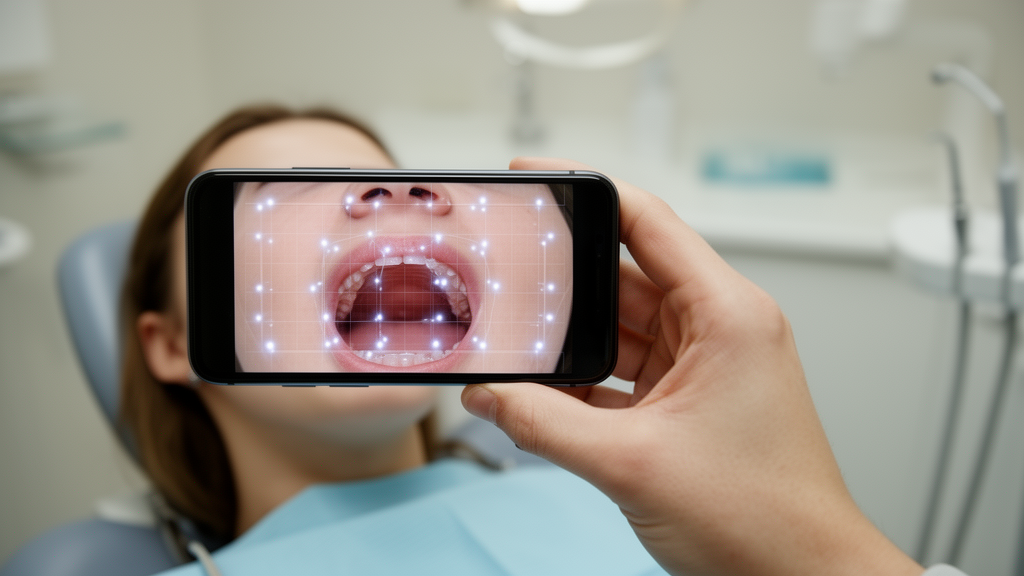
The dataset consisted of 240 palatal rugae images from healthy volunteers aged 18 to 40 years, recruited from an outpatient department, with 120 males and 120 females. Images were captured by trained personnel using a variety of commercially available smartphones with different manufacturers, sensor sizes and focal lengths, deliberately reflecting realistic field conditions. Each subject provided a single frontal intra-oral photograph of the anterior palate, taken with the participant seated upright, the mouth opened to expose the hard palate, the camera held roughly 15 to 25 centimetres away and aligned approximately perpendicular to the palatal plane. Ethical approval was obtained from an Institutional Ethics Committee following the Declaration of Helsinki, and written informed consent was collected from every participant.
Preprocessing was deliberately simple. Blurred images were discarded, the region of interest was manually cropped to isolate the rugae by a trained operator and verified by a senior clinician, and images were resized to 224 by 224 pixels with pixel values scaled to the range zero to one. No automated segmentation was used. Each image was labelled with the subject’s self-reported biological sex, and a second reviewer randomly inspected 20 percent of images for label accuracy and region quality, finding no systematic discrepancies. Data augmentation, including rotation, flipping and contrast and brightness adjustments, was applied during training to improve generalisation. The dataset was split 80 percent for training and 20 percent for testing, and all models ran on PyTorch using an Intel Core i7 processor, 16 gigabytes of RAM and an NVIDIA GeForce RTX GPU with 4 gigabytes of video memory.
ResNet50 achieved the highest overall accuracy at 77 percent, followed by VGG16 at 73 percent, EfficientNet-B0 at 69 percent and DenseNet121 at 62 percent. ResNet50 showed particularly strong sensitivity for female samples, with a recall of 0.88 and an F1-score of 0.79, corresponding to a per-class accuracy of 87.5 percent for females, while male classification was more balanced but weaker, with a recall of 0.67, an F1-score of 0.74 and a per-class accuracy of 66.7 percent. VGG16 produced comparatively stable results across both classes, with F1-scores of 0.75 for females and 0.71 for males. EfficientNet-B0 performed better on female images, with a recall of 0.83, but struggled on males, reaching a recall of only 0.54, which the authors attribute to limited extraction of fine-scale rugae geometry.
DenseNet121, despite being the deepest and most densely connected of the four architectures, underperformed with an accuracy of 62 percent and a marked reduction in male recall to 0.46. The researchers suggest this likely resulted from feature redundancy and overfitting on texture patterns in a small dataset. Receiver operating characteristic analysis supported the ranking: ResNet50 achieved the highest area under the curve, confirming the strongest separability between male and female classes, while VGG16 and EfficientNet-B0 showed moderate but reliable AUC values and DenseNet121 exhibited the lowest. The authors argue that moderately deep residual architectures offer the best balance between model depth, gradient stability and generalisation when analysing subtle biological variation in limited datasets.
Interpretability was addressed with gradient-weighted class activation mapping, or Grad-CAM, which highlights the image regions most influential in a model’s decision. For correctly classified cases, the heatmaps concentrated on the central and posterior rugae regions, areas the authors describe as anatomically recognised sites of greater inter-individual variation and sexual dimorphism. Representative examples showed ResNet50 focusing on these zones for both a correctly classified male image, with a confidence of 0.74, and a correctly classified female image, with a confidence of 0.59. Misclassified cases, by contrast, displayed dispersed attention toward non-rugae areas, suggesting potential interference from lighting or mucosal reflection during smartphone imaging. The team interprets this as evidence that the networks learned biologically meaningful cues rather than incidental artifacts.
The higher classification accuracy for female images across all four architectures aligns with prior morphological findings that female rugae tend to show more uniform and curved patterns, whereas male rugae often display greater irregularity and branching, increasing variability within the male class. The authors also note that ResNet50’s residual connections, which mitigate the vanishing gradient problem, enabled effective feature extraction despite the dataset’s modest size, while VGG16 and EfficientNet-B0 offered a favourable trade-off between computational cost and accuracy for resource-limited hardware.
The researchers are careful to frame the approach as an adjunct rather than a replacement for established forensic methods. The dataset is moderate in size and drawn from a single population, which may limit generalisability. Biological sex was self-reported rather than independently verified, and the analysis was conducted on living individuals, so the findings may not directly apply to postmortem cases, in which rugae may deteriorate during advanced decomposition and severe thermal damage may require invasive access to the palate under ethical and legal constraints. Image acquisition across different smartphone models introduces natural variability in resolution and illumination, although the authors argue this reflects realistic field conditions, and the region of interest was delineated manually rather than automatically.
Even with those caveats, the study demonstrates a feasible, low-cost and non-invasive pipeline: a smartphone photograph of the palate, processed through a fine-tuned pretrained network, can support preliminary biological sex estimation. The authors propose that the methodology could be integrated into mobile forensic kits or clinical software for rapid field-level screening in low-resource or rural settings, with Grad-CAM visualisation allowing investigators to verify the model’s decision focus before reporting. Future work, they say, will focus on expanding the dataset across broader demographic groups, incorporating multimodal dental data, testing performance under varied real-world conditions, exploring federated or edge-AI frameworks for privacy-preserving analysis, and adding further explainable artificial intelligence methods to support clinical and forensic applicability.
Subject of Research: Transfer learning with convolutional neural networks for biological sex classification from smartphone-acquired palatal rugae images
Article Title: Smartphone-based biological sex identification from palatal rugae images: a comparative transfer learning study of pretrained convolutional neural networks
Article References: Hussain, R., Pardeshi, R., Zainab, H., Hugar, D., Sultana, A., & Moinuddin, K. (2026). Smartphone-based biological sex identification from palatal rugae images: a comparative transfer learning study of pretrained convolutional neural networks. Egyptian Journal of Forensic Sciences, 16(1), Article 93. https://doi.org/10.1186/s41935-026-00592-z
Image Credits: AI Generated
DOI: 10.1186/s41935-026-00592-z
Keywords: palatal rugae, forensic odontology, convolutional neural networks, transfer learning, ResNet50, biological sex classification, smartphone imaging, Grad-CAM, biometrics, deep learning, Smartphone-based, biological
Cite Scienmag News
Ophelia Keating. (October 28, 2026). AI Reads Palatal Rugae From Smartphone Photos to Identify Sex. Scienmag. https://scienmag.com/ai-reads-palatal-rugae-from-smartphone-photos-to-identify-sex/
Ophelia Keating. "AI Reads Palatal Rugae From Smartphone Photos to Identify Sex." Scienmag, 28 October 2026, https://scienmag.com/ai-reads-palatal-rugae-from-smartphone-photos-to-identify-sex/. Accessed 28 October 2026.
Ophelia Keating. "AI Reads Palatal Rugae From Smartphone Photos to Identify Sex." Scienmag. October 28, 2026. https://scienmag.com/ai-reads-palatal-rugae-from-smartphone-photos-to-identify-sex/