


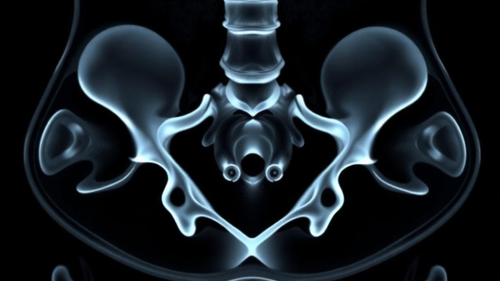
Every day, hospitals around the world generate millions of computed tomography scans, and most of them contain a quiet, underused treasure: images of the proximal femur, the upper end of the thigh bone that anchors the hip joint. A new nationwide study published in BMC Medical Imaging suggests that this routinely acquired imaging data can be mined automatically to reveal how bone structure changes with age and differs between men and women, potentially turning ordinary clinical scans into a large-scale screening tool for skeletal health. The research, led by Hanwen Cheng, Liwei Zhuang, Yiran Zhang and colleagues at Peking University People’s Hospital and collaborating institutions across China, demonstrates that machine learning can extract subtle, biologically meaningful patterns from hip CT images that the human eye routinely overlooks.
The study, registered retrospectively on ClinicalTrials.gov as NCT07162168, enrolled 4,152 participants who had undergone noncontrast routine CT scans. The cohort included 1,947 men, with a median age of 59 years spanning a remarkable range from 18 to 99. Rather than relying on slow, labor-intensive manual tracing of the femur, the team trained a deep learning segmentation model based on the nnU-Net framework, a self-configuring architecture widely regarded as one of the most robust tools for biomedical image segmentation. A subset of proximal femurs was first annotated by hand to teach the model exactly where the bone begins and ends in each volumetric scan. Once trained, the automated system could carve out the femoral volume of interest on its own, achieving a Dice similarity coefficient of 0.963 on the test set, a score indicating near-perfect overlap with expert annotations.
With the femur reliably isolated in thousands of scans, the researchers turned to radiomics, the practice of converting medical images into hundreds of quantitative features that describe texture, shape, intensity and spatial patterns within a defined region. Using the open-source PyRadiomics package, they extracted features from each segmented femur, following standardized conventions designed to make radiomic measurements reproducible across scanners and institutions. The idea behind radiomics is deceptively simple: the microscopic architecture of bone, including trabecular thinning, cortical porosity and the redistribution of mineral, leaves fingerprints in the statistical texture of a CT image. Those fingerprints, aggregated across thousands of features, may encode a person’s skeletal age far more richly than a single density measurement.
To test whether these features actually carry information about aging and sex, the team built machine learning models in a development cohort of 2,969 participants and then evaluated them in a completely separate external validation cohort of 1,183 participants, a design that guards against the optimistic results that plague many single-center AI studies. For age, the best performer was a support vector regression model, which estimated chronological age from femoral radiomics with a coefficient of determination of 0.50 in external validation, a mean absolute error of 8.84 years and a root mean squared error of 11.32 years. In plain terms, the model could roughly halve the uncertainty of a blind guess about a person’s age using nothing but the texture of their hip bone.
Sex classification proved even more striking. Two algorithms, support vector classification and the gradient-boosting method XGBoost, both achieved an area under the receiver operating characteristic curve of 0.94 in external validation, with support vector classification showing slightly higher accuracy and F1-score. An AUC of 0.94 means the model distinguished male from female skeletons with high reliability across the entire cohort, capturing structural sexual dimorphism in the femur that goes well beyond simple size differences. Because the features were extracted automatically from routine scans, the result hints that sex-related skeletal architecture is legible in ordinary clinical imaging without any dedicated bone protocol.
Perhaps the most consequential finding concerns the relationship between the radiomics-derived age index and actual bone mineral biology. In the external validation cohort, the researchers compared their radiomic age index against volumetric bone mineral density of the femoral neck measured by quantitative computed tomography, the gold-standard three-dimensional density technique. The radiomics index tracked vBMD more strongly than chronological age did, with a Spearman correlation of minus 0.546 versus minus 0.443 for age alone, a difference the authors confirmed statistically using Steiger’s test (Z = 4.97, P < 0.001). This comparison matters because it suggests the radiomic signature is not merely a proxy for how many birthdays a person has had, but a reflection of genuine structural deterioration in the bone, the kind of change that underlies osteoporosis and hip fracture risk.
The clinical logic here is what researchers call opportunistic imaging. Dual-energy X-ray absorptiometry, the standard test for osteoporosis, is underused: many people at risk never get scanned, and fractures often arrive as the first sign of disease. But a large fraction of older adults undergo abdominal, chest or pelvic CT for unrelated reasons, and those scans routinely capture the proximal femur. If an automated pipeline can convert that incidental imaging into a skeletal age index or a bone quality flag, clinicians could identify patients with accelerated bone aging who would benefit from confirmatory density testing or preventive treatment, all without a single additional scan, extra radiation dose or added visit. The authors caution that their radiomic patterns still need validation against hard clinical outcomes such as incident fractures before such screening becomes practice, but the biological anchoring to vBMD is an encouraging first step.
The technical achievement should not be undersold either. Automated segmentation at a Dice coefficient above 0.96 removes the bottleneck that has historically limited radiomics to small, single-center studies requiring hours of expert contouring. By combining nnU-Net segmentation, standardized PyRadiomics extraction and rigorous external validation across nationwide cohorts, the study offers a template for how imaging biomarkers can be scaled to population level. The work was supported by the Beijing Natural Science Foundation-Fengtai Joint Innovation Project, the National Natural Science Foundation of China Science Center Program and the Shenzhen Medical Research Fund, and it was approved by the ethics committee of Peking University People’s Hospital with informed consent waived for the retrospective use of de-identified images.
There are, of course, caveats to keep in mind. The cohort was drawn from Chinese hospitals, and radiomic features can be sensitive to scanner vendor, reconstruction settings and dose, so multicenter generalization beyond this dataset remains to be demonstrated. The age model’s mean absolute error of nearly nine years is impressive for image-only prediction but far too coarse to serve as a precise biological clock for any individual. And the study measured associations with density, not with fractures, falls or mortality, so the ultimate clinical value hinges on longitudinal follow-up. Still, the direction of travel is clear: the femur that appears incidentally on a routine scan carries a quantifiable structural biography, written in texture patterns that machines can now read at scale.
If subsequent studies confirm that radiomics-derived bone age predicts fractures and responds to osteoporosis treatment, the implications could extend well beyond orthopedics. Bone is a living archive of hormonal change, nutrition, inflammation and mechanical loading, and a cheap, automated readout of its microarchitecture could inform research on aging itself, from sarcopenia to metabolic bone disease. For now, the study stands as a proof of concept that the skeleton’s quiet remodeling, normally invisible in the reading room, can be decoded from data that hospitals already possess, waiting in the archive.
Subject of Research: Automated CT radiomics of the proximal femur for quantifying age- and sex-related bone structural changes
Article Title: Automated proximal femur CT radiomics identifies age- and sex-related radiomic patterns of bone structure: a nationwide multicohort validation study
Article References: Cheng, H., Zhuang, L., Zhang, Y., Ran, Y., Jiang, B., & Kou, Y. (2026). Automated proximal femur CT radiomics identifies age- and sex-related radiomic patterns of bone structure: a nationwide multicohort validation study. BMC Medical Imaging. https://doi.org/10.1186/s12880-026-02836-9
Image Credits: AI Generated
DOI: 10.1186/s12880-026-02836-9
Keywords: radiomics, proximal femur, CT imaging, deep learning, nnU-Net, bone aging, bone mineral density, osteoporosis, machine learning, opportunistic screening, quantitative CT, hip fracture risk
Cite Scienmag News
Beatrice Stafford. (September 30, 2026). AI Reads Hidden Bone Aging Patterns in Routine CT Scans of the Hip. Scienmag. https://scienmag.com/ai-reads-hidden-bone-aging-patterns-in-routine-ct-scans-of-the-hip/
Beatrice Stafford. "AI Reads Hidden Bone Aging Patterns in Routine CT Scans of the Hip." Scienmag, 30 September 2026, https://scienmag.com/ai-reads-hidden-bone-aging-patterns-in-routine-ct-scans-of-the-hip/. Accessed 4 October 2026.
Beatrice Stafford. "AI Reads Hidden Bone Aging Patterns in Routine CT Scans of the Hip." Scienmag. September 30, 2026. https://scienmag.com/ai-reads-hidden-bone-aging-patterns-in-routine-ct-scans-of-the-hip/


