


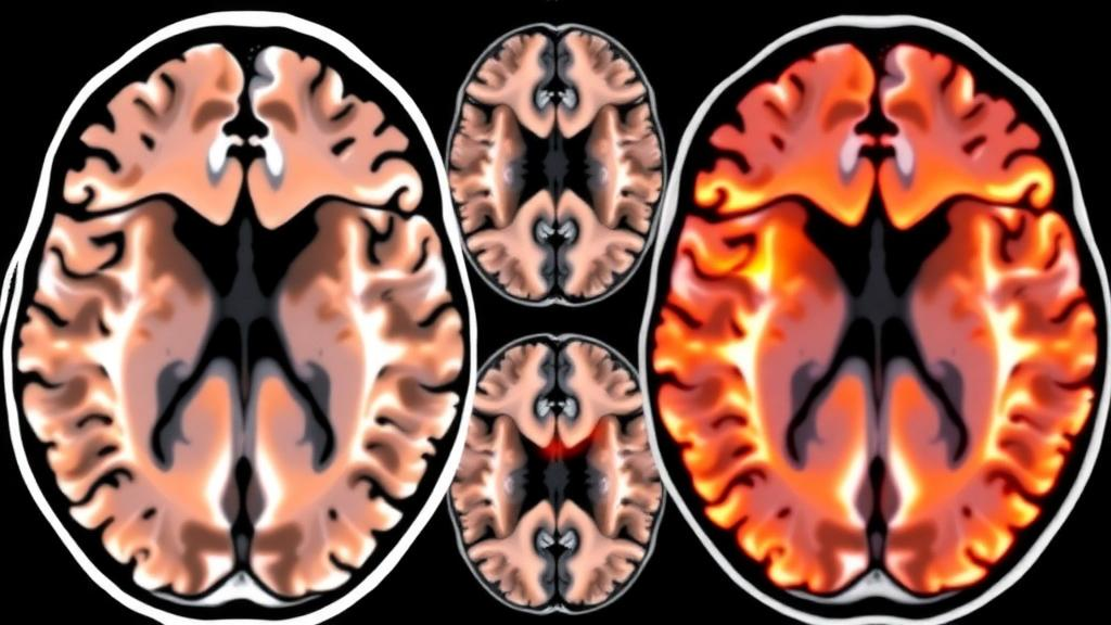
When a patient arrives at a hospital with suspected stroke, every minute of delay means more brain tissue lost. Physicians often turn first to magnetic resonance imaging sequences that do not require an injected contrast dye, because these scans are faster to obtain and more widely available in everyday clinical settings than their contrast-enhanced counterparts. The most common of these, the FLAIR sequence, suppresses the bright signal of cerebrospinal fluid so that damaged tissue stands out more clearly to a trained radiologist. Yet the very properties that make contrast-free imaging convenient in the emergency room also make it notoriously difficult for computer programs to read automatically. The damaged regions frequently appear faint, their shapes are irregular and unpredictable, and their appearance can shift noticeably from one MRI machine to the next. A new peer-reviewed study, published ahead of print in the Journal of Intelligent Systems in Current Computer Engineering, set out to measure exactly how well modern artificial intelligence can overcome these obstacles.
The research team, led by Patricia García-Berlanga and including Juan Zapata, Juan Martinez-Alajarin, and Pablo Hernandez-Cerdan, compared three distinct families of deep learning models for detecting stroke lesions on non-contrast MRI. Their motivation is straightforward: interpreting these scans currently depends heavily on the judgment of a radiologist, and automating even part of that process, as a support tool rather than a replacement, could accelerate triage and reduce the variability that naturally arises between different human readers. Building reliable models for FLAIR images has proven stubbornly difficult, however, precisely because of the features that make these scans attractive in the first place. The contrast between healthy and injured tissue is lower than in contrast-enhanced imaging, and the images can look markedly different depending on the scanner model or the hospital that produced them. Any automated system must therefore be robust not only to the subtle appearance of the lesions themselves but also to the considerable technical variation across imaging hardware.
To make the comparison meaningful, the researchers evaluated three well-known image-segmentation architectures under strictly matched conditions. The first was a classical three-dimensional U-Net, a manually tuned convolutional neural network that has served as a workhorse of medical image segmentation for years. The U-Net design uses an encoder that compresses an image into increasingly abstract feature maps and a decoder that expands those features back into a full-resolution prediction, with skip connections that preserve fine spatial detail. The second contender was nnU-Net, a newer self-configuring framework that automatically adapts its preprocessing, architecture, and training schedule to the specific dataset it is given, dramatically reducing the need for laborious manual trial and error. The third was an adapted version of YOLOv8, an object-detection architecture better known for real-time applications such as video analysis, which the authors modified to perform two-dimensional segmentation of MRI slices.
All three models were trained and tested on the ISLES 2022 dataset, a publicly available benchmark of 250 stroke MRI cases that is widely used in the lesion-segmentation community. Crucially, the same preprocessing pipeline and the same evaluation procedures were applied to every model, ensuring that differences in performance could be attributed to the architectures themselves rather than to unequal treatment. Performance was quantified with two standard metrics for segmentation accuracy: the Dice similarity coefficient and the Intersection over Union, commonly abbreviated IoU. Both metrics measure how closely the AI’s predicted lesion boundary overlaps the lesion as delineated by expert annotators, with a score of one indicating perfect agreement and a score of zero indicating no overlap at all. Because these metrics penalize both missed tissue and falsely labeled healthy tissue, they provide a balanced picture of where each model excels and where it falters.
The results delivered a clear verdict on accuracy. The self-configuring nnU-Net came out on top, achieving the highest average overlap with expert-marked lesions, with a Dice score of approximately 0.483. While that number may appear modest on an absolute scale, it reflects the genuine difficulty of the task on contrast-free images, where lesion boundaries are ambiguous even for human experts. Importantly, the nnU-Net held up comparatively well across the wide variety of lesion shapes and sizes represented in the dataset, suggesting that its automatic configuration strategy successfully generalizes to the heterogeneity that characterizes real clinical material. The framework’s ability to tune itself to a new dataset, rather than relying on hand-crafted settings, appears to be the decisive advantage in this setting.
The adapted YOLOv8 model was not far behind on precision, posting a Dice score of around 0.476, a difference small enough that the two leading approaches are functionally comparable in terms of overlap quality. What distinguished YOLOv8 was speed. It processed scans substantially faster than the other two methods, and the authors suggest that this could make it a strong candidate for time-sensitive clinical workflows where throughput matters as much as raw accuracy. In an acute stroke setting, where imaging findings must reach the treating physician within minutes, a model that sacrifices a sliver of precision for a large gain in processing time may ultimately deliver more clinical value than a slower but marginally more accurate alternative. This speed-accuracy trade-off is a recurring theme in deployed medical AI systems, and the study provides concrete numbers to inform that decision.
The classical 3D U-Net, included as a manually configured baseline, performed noticeably worse overall, with a Dice score of roughly 0.294. The researchers attribute this gap to the architecture’s greater sensitivity to manual parameter choices and to variation across the dataset. In other words, when the settings are chosen by hand rather than learned automatically, small suboptimal decisions can compound into substantially degraded performance, particularly when the training data span a broad range of lesion appearances and scanner characteristics. The result is a cautionary tale for research groups still relying on fully manual pipelines: the additional engineering effort of self-configuring frameworks appears to pay measurable dividends on challenging, heterogeneous clinical data.
Beyond the headline numbers, the study dug into the specific failure modes shared by all three approaches. Common trouble spots included small lesions located near the outer edges of the brain, where anatomical complexity and partial-volume effects confound the models. Diffuse, low-contrast regions of damage also proved inherently hard to distinguish from healthy tissue, since the signal differences that define such lesions sit close to the noise floor of the image. A third recurring problem was spatial misalignment, in which the predicted lesion outline did not correspond well to the actual location of the abnormality within the scan. The authors also identified preprocessing techniques that helped mitigate these weaknesses, including resampling to standardize the voxel spacing across scans, normalizing pixel intensity values so that images from different scanners share a common scale, and training the networks on smaller image patches rather than whole volumes at once, which increases the effective diversity of training examples and focuses learning on local texture.
From these findings, the researchers distilled practical recommendations for teams building similar tools. Where computing resources permit, self-configuring frameworks such as nnU-Net are worth prioritizing for their robustness and reduced dependence on expert tuning. In environments where processing speed is the binding constraint, lightweight architectures in the mold of the adapted YOLOv8 represent a reasonable and defensible trade-off. Regardless of which architecture is chosen, the authors stress that rigorous validation across multiple clinical sites and scanner types is essential before any such tool can responsibly be considered for real-world clinical use, given how profoundly scanner-to-scanner variability can affect performance. A model that excels on a single institutional dataset may falter on images from an unfamiliar machine, and only multi-site testing can reveal whether a system has genuinely learned the imaging signature of stroke or merely the quirks of one scanner population.
The study, titled Detection of Vascular Pathologies in Non-contrast Magnetic Resonance Images for Early Screening, contributes a carefully controlled benchmark that clinicians and engineers alike can use to calibrate expectations for automated stroke screening. Its central message is encouraging: modern self-configuring and lightweight deep learning models can now extract clinically meaningful lesion information from the fast, contrast-free MRI sequences that emergency departments already rely on, without the delays and costs of contrast administration. At the same time, the modest absolute Dice scores and the catalog of shared failure modes make clear that these systems remain support tools rather than autonomous diagnosticians. The path forward, the authors suggest, lies in combining the accuracy of self-configuring frameworks with the speed of streamlined detectors, validated across the messy diversity of real-world scanners, so that automated lesion detection can one day help shave precious minutes off the interval between a patient’s arrival and a life-saving treatment decision.
Subject of Research: Deep learning segmentation of stroke lesions on non-contrast FLAIR MRI scans
Article Title: AI models put to the test for spotting stroke damage on routine brain scans
Article References: AI models put to the test for spotting stroke damage on routine brain scans. (n.d.). Original publication
Image Credits: AI Generated
DOI: Not provided
Keywords: artificial intelligence, deep learning, stroke, MRI, FLAIR, nnU-Net, YOLOv8, U-Net, image segmentation, ISLES 2022, medical imaging, clinical decision support
Cite Scienmag News
Cassandra Pierce. (October 4, 2026). AI Models Face Off in Head-to-Head Test for Spotting Stroke Damage on Routine Brain Scans. Scienmag. https://scienmag.com/ai-models-face-off-in-head-to-head-test-for-spotting-stroke-damage-on-routine-brain-scans/
Cassandra Pierce. "AI Models Face Off in Head-to-Head Test for Spotting Stroke Damage on Routine Brain Scans." Scienmag, 4 October 2026, https://scienmag.com/ai-models-face-off-in-head-to-head-test-for-spotting-stroke-damage-on-routine-brain-scans/. Accessed 4 October 2026.
Cassandra Pierce. "AI Models Face Off in Head-to-Head Test for Spotting Stroke Damage on Routine Brain Scans." Scienmag. October 4, 2026. https://scienmag.com/ai-models-face-off-in-head-to-head-test-for-spotting-stroke-damage-on-routine-brain-scans/


