


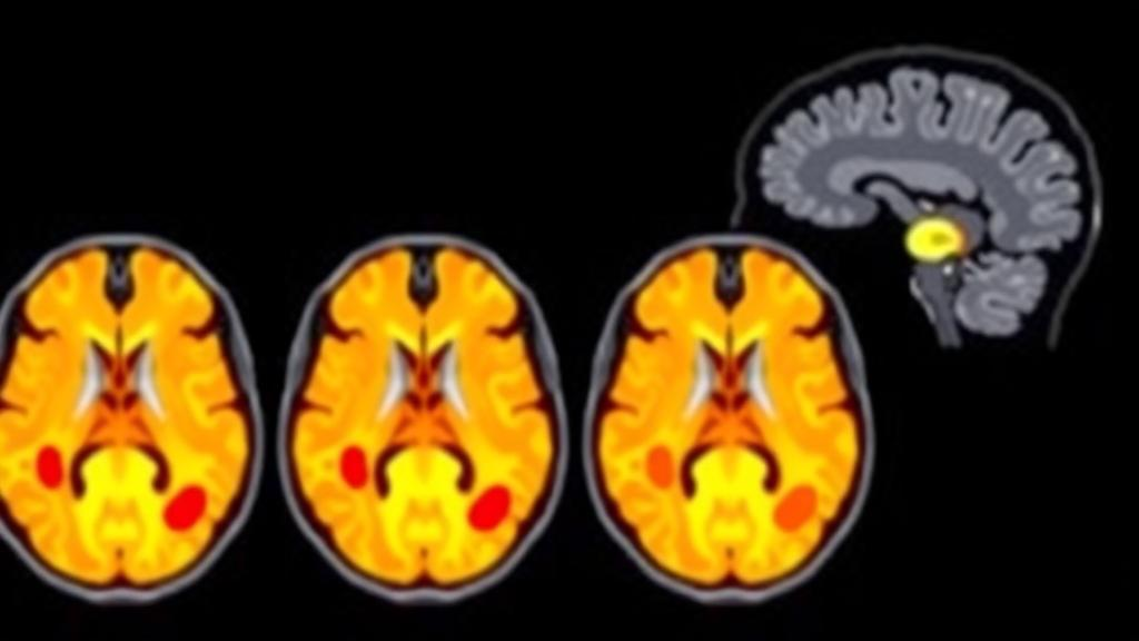
Multiple sclerosis is one of the most common chronic neurological disorders in the world, a condition in which the immune system strips away the protective myelin sheath surrounding nerve fibers in the central nervous system. The resulting lesions disrupt the transmission of electrical impulses and gradually erode motor control, sensation, and vision. Because the disease can smolder silently for years before symptoms become disabling, catching lesions early and measuring them precisely is one of the most valuable things modern medicine can do for patients. Now, a team of researchers from United International University in Dhaka and Charles Darwin University in Australia has unveiled a deep learning framework called MedSegNet-AXU that not only detects and outlines these lesions with remarkable precision but also turns them into a window on how the disease affects different body systems. The work, published open access in Complex & Intelligent Systems, describes a Dice score of 98.58 percent for lesion segmentation, a specificity of 98.71 percent, and a graph-based classification pipeline that predicts functional impairment with accuracies reaching 96.55 percent.
The heart of the new system is a carefully engineered convolutional neural network built on the U-Net architecture, a design that has become the workhorse of medical image segmentation since its introduction. U-Net’s signature shape, an encoder that compresses an image into abstract features followed by a decoder that expands those features back into a pixel-level mask, is well suited to outlining structures in three-dimensional brain scans. But standard U-Net models often struggle with the peculiar challenges of multiple sclerosis lesions, which are small, irregularly shaped, scattered throughout the white matter, and easily confused with other bright spots on magnetic resonance images. The research team addressed this by bolting two powerful attention mechanisms onto the backbone. The first is the Convolutional Block Attention Module, known as CBAM, which refines feature maps along two complementary axes: it learns which channels of information matter most and which spatial regions deserve focus, effectively teaching the network where to look and what to weigh.
The second enhancement is an Extended Atrous Spatial Pyramid Pooling module, a technique borrowed from semantic segmentation research that solves a different problem: scale. Lesions in the brain come in wildly different sizes, from a few voxels to sprawling patches of demyelination, and a network that only looks at one resolution tends to miss the extremes. Atrous convolution, sometimes called dilated convolution, widens the receptive field of the filters without adding parameters or losing resolution, allowing the model to sample context at multiple scales simultaneously. By stacking these dilated convolutions in a pyramid arrangement, the module captures both the fine texture of a tiny lesion and the broader anatomical context of a large one. The combination of channel-wise attention, spatial attention, and multi-scale context extraction is what the authors credit for the model’s standout performance on the Brain Magnetic Resonance Dataset of Multiple Sclerosis, where it achieved its near-perfect Dice score, a metric that measures the overlap between the machine’s segmentation and expert annotations.
Impressive as those numbers are, the team did not stop at a single dataset. To test whether the architecture had genuinely learned the anatomy of abnormal tissue rather than memorizing the quirks of one collection of scans, they evaluated MedSegNet-AXU on the widely used Brain Tumor Segmentation challenges from 2019, 2020, and 2021. Across different imaging modalities and tumor sub-regions, the model delivered mean Dice scores between 92 and 96 percent and Jaccard indices, a stricter measure of overlap, between 85 and 91 percent. This cross-domain validation matters because a segmentation tool that only works on one dataset is a laboratory curiosity, while one that generalizes across diseases and imaging protocols could become a genuine clinical instrument. The results suggest that the attention-driven design captures features of pathological tissue that transcend the specific appearance of multiple sclerosis lesions.
But segmentation, however accurate, is only the first half of the story. Once the network had traced the boundaries of each lesion, the researchers extracted 21 radiomic features from the segmented tissue, quantitative descriptors that capture properties such as shape, intensity distribution, and texture that the human eye cannot reliably grade. Crucially, they organized this analysis around three functional systems that multiple sclerosis attacks: the sensory, motor, and visual pathways. The idea is elegant in its simplicity. If the mathematical fingerprint of a lesion correlates with the symptoms a patient experiences, then a scan alone could hint at how the disease is manifesting, potentially flagging damage before a clinical exam reveals it. The radiomic features were then fused with clinical metadata, information about the patients themselves, to create a richer description of each case than imaging or demographics could provide alone.
With dozens of candidate features in hand, the team faced the classic machine learning dilemma of separating signal from noise. They applied Chi-square feature selection, a statistical test that scores each feature by how strongly its distribution relates to the outcome of interest, and retained the top 20 for classification. Then came the most novel step. Rather than feeding these features into conventional classifiers alone, the researchers represented each patient as a node in a graph, connected by edges that encode relationships in the data, and trained graph neural networks to make predictions. Among the models tested, GraphSAGE stood out dramatically. This algorithm works by allowing each node to aggregate information from its neighbors, layer by layer, so that a patient’s classification is informed not just by their own features but by those of similar patients in the learned graph structure. It achieved test accuracies of 86.21 percent for the sensory system, 96.55 percent for the motor system, and 75.86 percent for the visual system, significantly outperforming the traditional machine learning baselines.
High accuracy alone can be misleading in medical machine learning, where a model may latch onto spurious correlations that vanish in the clinic. The researchers therefore subjected GraphSAGE to two independent reliability checks. The first, Threshold Analysis with Anomaly Edges, probes how the model behaves when unusual or out-of-distribution connections appear in the graph, testing whether its predictions degrade gracefully or collapse when faced with atypical patients. The second, Correlation Range Clustering, examines the structure of the relationships the model relies on, verifying that the graph edges reflect meaningful similarity rather than artifacts of construction. The fact that the model survived these stress tests lends credibility to the headline numbers and addresses one of the most persistent criticisms of deep learning in medicine: that its predictions are often opaque and fragile in the face of real-world variability.
The clinical implications of this pipeline are considerable. Today, diagnosing and monitoring multiple sclerosis depends on radiologists manually counting and measuring lesions across time points, a laborious process prone to inter-observer disagreement, and on clinical scales that quantify disability through examination. A framework that automatically segments lesions, extracts quantitative features, and links them to functional systems could standardize this workflow, reduce the variability that plagues longitudinal monitoring, and give neurologists an earlier, more objective read on disease activity. The fusion of imaging with clinical metadata also points toward personalized medicine: if a patient’s lesion profile predicts motor decline, for example, treatment intensity might be adjusted before irreversible damage accumulates. The authors frame the approach as a comprehensive tool for lesion analysis aimed at precise diagnosis and personalized treatment strategies, and the architecture’s demonstrated portability to tumor segmentation hints at applications well beyond one disease.
Caveats remain, as they always do at this stage of translation. The model was trained and validated on research datasets rather than deployed in a live hospital setting, and the classification accuracies, while strong, vary across the three functional systems, with the visual system proving hardest to predict. Prospective studies in diverse patient populations will be needed to confirm that the performance holds under the messier conditions of routine clinical imaging, with different scanners, protocols, and artifact levels. Nevertheless, the study represents a notable convergence of several trends in medical artificial intelligence: attention mechanisms that make segmentation networks more discerning, radiomics that convert images into measurable biology, and graph learning that exploits the relationships among patients rather than treating each case in isolation. Published open access with a permanent DOI, the work invites other groups to build on it, and it offers a glimpse of a future in which a routine brain scan quietly tells a neurologist not just where the lesions are, but what they mean for the person carrying them.
Subject of Research: Deep learning segmentation of multiple sclerosis lesions and graph-based classification using MRI radiomics and clinical data
Article Title: MedSegNet-AXU: advanced segmentation and graph-based classification of multiple sclerosis lesions from 3D magnetic resonance imaging via radiomics and clinical data fusion
Article References: Karim, W., Zaman, S. B., Sutradhar, D., Debnath, R. K., Azam, S., Yeo, K. C., Zhang, Y., & Jonkman, M. (2026). MedSegNet-AXU: advanced segmentation and graph-based classification of multiple sclerosis lesions from 3D magnetic resonance imaging via radiomics and clinical data fusion. Complex & Intelligent Systems. https://doi.org/10.1007/s40747-026-02535-6
Image Credits: AI Generated
DOI: 10.1007/s40747-026-02535-6
Keywords: multiple sclerosis, lesion segmentation, 3D brain MRI, U-Net, attention mechanism, CBAM, atrous spatial pyramid pooling, radiomics, graph neural networks, GraphSAGE, feature selection, clinical data fusion
Cite Scienmag News
Cassandra Pierce. (October 8, 2026). AI Framework Spots MS Lesions With Record Accuracy by Fusing MRI and Clinical Data. Scienmag. https://scienmag.com/ai-framework-spots-ms-lesions-with-record-accuracy-by-fusing-mri-and-clinical-data/
Cassandra Pierce. "AI Framework Spots MS Lesions With Record Accuracy by Fusing MRI and Clinical Data." Scienmag, 8 October 2026, https://scienmag.com/ai-framework-spots-ms-lesions-with-record-accuracy-by-fusing-mri-and-clinical-data/. Accessed 8 October 2026.
Cassandra Pierce. "AI Framework Spots MS Lesions With Record Accuracy by Fusing MRI and Clinical Data." Scienmag. October 8, 2026. https://scienmag.com/ai-framework-spots-ms-lesions-with-record-accuracy-by-fusing-mri-and-clinical-data/


