


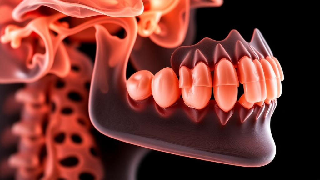
Deep inside the upper neck, just below the skull, sits a small, peg-shaped bone that forensic scientists have long suspected holds clues to a person’s biological sex. Now, a new study published in the International Journal of Legal Medicine suggests that this bone, the odontoid process of the second cervical vertebra, may carry far more information than its modest size implies. Using a form of artificial intelligence applied to dental cone-beam computed tomography scans, researchers in Istanbul have shown that quantitative image features extracted from this structure can distinguish male from female skeletons with impressive internal accuracy, potentially offering a new tool for identifying human remains when only fragments of the skeleton are available.
The research, led by Alperen Tekin and colleagues at Istanbul Medeniyet University, addresses a persistent challenge in forensic anthropology. When investigators recover skeletal remains, estimating sex is one of the first and most important steps, because it narrows the pool of missing persons and shapes every subsequent analysis of age, stature, and ancestry. The most reliable methods rely on the pelvis and skull, but these bones are frequently missing, fragmented, or damaged in disaster scenes, fires, and archaeological contexts. Vertebrae, which are dense and often survive, have therefore attracted growing attention as alternative sex indicators, and the second cervical vertebra, known as the axis or C2, has emerged as a particularly promising candidate.
What makes the new study distinctive is its source of imaging data. Cone-beam computed tomography, or CBCT, is a low-dose scanning technology used routinely in dentistry and maxillofacial radiology, and the axis vertebra falls within the field of view of many head-and-neck CBCT examinations. This means that vast archives of existing clinical scans, acquired for entirely unrelated dental reasons, could in principle be mined for forensic information without exposing anyone to additional radiation. The researchers exploited this opportunity by retrospectively analyzing 140 adult CBCT examinations, 68 from female patients and 72 from male patients, all acquired on a single scanner under homogeneous imaging conditions.
Rather than measuring the bone manually with calipers, as traditional morphometric studies do, the team turned to radiomics, a computational approach that converts medical images into hundreds of quantitative features. Radiomics captures characteristics that the human eye cannot reliably assess, including the statistical distribution of gray values, the coarseness and uniformity of bone texture, and the precise three-dimensional shape of the segmented structure. From each scan, the researchers extracted 107 original-image radiomic features from two different regions of interest: the entire second cervical vertebra, and the odontoid process alone, the tooth-like projection that rises from the vertebral body and allows the head to rotate.
The central question was methodological as much as forensic. If a forensic team only has access to the odontoid process, perhaps because the rest of the vertebra is damaged or outside the scan volume, does segmenting just that smaller structure sacrifice the discriminative information contained in the whole bone? To find out, the researchers built paired prediction models for each region of interest, using regularized machine learning algorithms, including least absolute shrinkage and selection operator, or LASSO, regression and elastic-net regression, both of which are designed to handle datasets where the number of features is large relative to the number of samples.
The results, validated through a rigorous internal testing framework, were strikingly consistent. Across 100 repeated paired validations with a balanced 80/20 train-test split, the mean area under the receiver operating characteristic curve, or AUC, reached 0.929 for the combined whole-C2 models, while the odontoid-only models achieved mean AUCs of 0.924 for texture-only features, 0.918 for combined LASSO models, and 0.905 for combined elastic-net models. An AUC of 0.5 would indicate performance no better than a coin flip, while 1.0 represents perfect discrimination, so values above 0.9 indicate strong internal classification ability. Five-fold nested cross-validation, a more conservative scheme that guards against information leaking from the test data into model training, produced AUCs of 0.918 for the whole-C2 combined LASSO model and 0.914 for the odontoid-only equivalent, confirming that the advantage of the whole vertebra was marginal at best.
The team also took deliberate steps to guard against the inflated performance estimates that have plagued much of the radiomics literature. All preprocessing steps were fitted exclusively on the training set, label-permutation testing was used to verify that the models were genuinely learning signal rather than noise, and the permutation null distributions were centered near 0.50, with a permutation p-value of 0.003 for both regions of interest. The authors were candid about the limits of their most eye-catching numbers: the highest AUCs observed on the primary test split, which ranged from 0.985 to 0.995, were derived from only 28 test subjects and were explicitly interpreted as optimistic estimates rather than realistic expectations of casework performance.
Perhaps the most consequential finding is that the odontoid process alone, a structure smaller than a fingertip, carries nearly as much sex-related information as the entire vertebra. The whole-C2 region retained more shape information, which makes anatomical sense given that the axis vertebra shows well-documented sexual dimorphism in its overall dimensions, but the texture features computed from the odontoid process alone performed on a par with the full-bone models. For forensic practice, this matters because the odontoid process is compact, anatomically well-defined, and easy to segment, and because it may be the only part of the vertebra preserved or captured in certain imaging scenarios. Previous studies had already shown that odontoid volume and morphometry differ between sexes, and the new results suggest that subtle internal bone texture adds a further layer of discriminative signal.
At the same time, the researchers are careful not to oversell the approach, and their caveats deserve as much attention as their headline numbers. The study was retrospective and single-device, meaning all scans came from one scanner and one institution under homogeneous imaging conditions, so the models have not yet faced the variability of different machines, scanning protocols, and populations. No external validation was performed, the reliability subsets used to assess feature reproducibility were too small to support comparative conclusions between the two regions of interest, and the biological meaning of many texture descriptors remains uncertain. High dimensionality and possible instability in feature selection further complicate interpretation, and the authors note that these limitations restrict the applicability of the models to real forensic casework for now.
Even with those caveats, the study represents a meaningful step toward a future in which routine dental scans, already acquired for millions of patients, double as a forensic resource. The work aligns with international standardization efforts such as the Image Biomarker Standardization Initiative and follows modern reporting frameworks for prediction models, signaling a maturing field that is increasingly aware of its own methodological pitfalls. If future multi-center studies can confirm that C2 radiomics generalizes across scanners and populations, forensic investigators may one day load an anonymous CBCT scan into software, segment a bone the size of a thumb, and receive a statistically grounded estimate of sex within seconds, turning an everyday dental image into a silent witness that helps put a name to the nameless.
Subject of Research: CBCT radiomics of the second cervical vertebra for forensic sex estimation
Article Title: Whole-axis versus odontoid process CBCT radiomics for forensic sex estimation: a retrospective paired-ROI model-development study
Article References: Tekin, A., Deveci, B., Tekin, G., & Ay, F. (2026). Whole-axis versus odontoid process CBCT radiomics for forensic sex estimation: a retrospective paired-ROI model-development study. International Journal of Legal Medicine. https://doi.org/10.1007/s00414-026-03999-4
Image Credits: AI Generated
DOI: 10.1007/s00414-026-03999-4
Keywords: forensic anthropology, radiomics, cone-beam computed tomography, second cervical vertebra, odontoid process, sex estimation, machine learning, forensic radiology, LASSO, elastic net, bone texture analysis, International Journal of Legal Medicine
Cite Scienmag News
Ophelia Keating. (October 3, 2026). A Tiny Neck Bone May Reveal Your Sex: AI Reads Hidden Patterns in Dental Scans. Scienmag. https://scienmag.com/a-tiny-neck-bone-may-reveal-your-sex-ai-reads-hidden-patterns-in-dental-scans/
Ophelia Keating. "A Tiny Neck Bone May Reveal Your Sex: AI Reads Hidden Patterns in Dental Scans." Scienmag, 3 October 2026, https://scienmag.com/a-tiny-neck-bone-may-reveal-your-sex-ai-reads-hidden-patterns-in-dental-scans/. Accessed 4 October 2026.
Ophelia Keating. "A Tiny Neck Bone May Reveal Your Sex: AI Reads Hidden Patterns in Dental Scans." Scienmag. October 3, 2026. https://scienmag.com/a-tiny-neck-bone-may-reveal-your-sex-ai-reads-hidden-patterns-in-dental-scans/


