


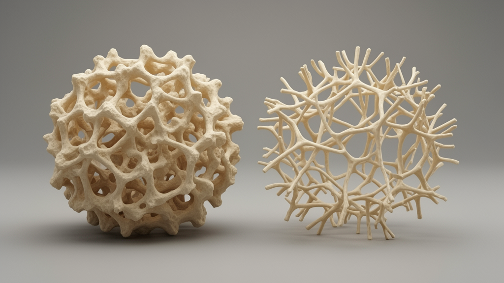
Deep inside the hip, a quiet architectural collapse precedes the loud crack of an osteoporotic fracture. A research team in China has now documented that collapse in remarkable detail, showing that the spongy trabecular bone of the femoral head in patients who fractured their femoral neck is not merely less dense than normal bone but structurally and mechanically devastated. The study, published in Frontiers in Medicine, combined high-resolution micro-CT imaging with direct laboratory strength testing of bone specimens, and found that the degree of microstructural damage tracks closely with the loss of mechanical performance. The work offers some of the most tangible evidence yet that bone quality, not just bone quantity, determines whether a fall from standing height ends in a shattered hip.
Osteoporosis is a systemic skeletal disease defined by low bone mass, deterioration of bone microarchitecture, and increased fragility, and it is often called a silent killer because it progresses without symptoms until a vertebra or hip gives way. Epidemiological surveys in China report age-standardized prevalence rates among people over 50 of 6.46 percent for men and 29.13 percent for women, and the burden grows as populations age. Clinically, the diagnostic gold standard remains bone mineral density testing, but density alone has relatively low sensitivity and specificity for predicting fracture risk. Bone strength depends not only on how much mineral the tissue contains but also on the geometry and connectivity of its internal trabecular scaffold, features that are difficult to assess in a living patient. That gap between what scans measure and what bone can withstand is precisely what the new study set out to probe.
The researchers, led by Hao Liu and Zhizheng Xiong of Yueyang Hospital Affiliated to Hunan Normal University and Yueyang People’s Hospital, with colleagues from Xi’an Jiaotong University, obtained femoral heads discarded after hip replacement surgery. The experimental group comprised 20 patients who had suffered osteoporotic femoral neck fractures after low-energy trauma such as a fall from standing height, aged 69 to 90 years with a mean of 80.65 years. The control group consisted of 13 patients undergoing replacement for early-stage degenerative hip disease, aged 49 to 83 years with a mean of 64.08 years. Patients with bone tumors, rheumatoid arthritis, ankylosing spondylitis, or femoral head necrosis were excluded. All specimens were preserved in 10 percent neutral buffered formalin and processed through identical harvesting, scanning, and testing workflows, with sampling performed by a single operator following a unified standard operating procedure.
Specimen preparation was deliberately standardized. After micro-CT scanning of each intact femoral head, the neck was divided into three equal parts from front to back, and only the middle slice was sampled. Using a hollow saw, the team extracted cylindrical cancellous bone plugs 8 millimeters in diameter from the zone of maximal diameter, oriented along the principal compressive trabeculae, the load-bearing struts that follow the stress trajectories of the femoral head. Marking the sampling location and orientation on each specimen before cutting minimized variability. This approach ensured that every mechanical test interrogated bone from the same anatomical region and directional framework in both groups, reducing systematic sampling bias that could otherwise masquerade as disease-related differences.
Imaging was performed with a SkyScan 1276 micro-CT scanner at a voxel resolution of approximately 36 micrometers, using a 100 kilovolt tube voltage, 200 microampere current, 211 millisecond exposure, and a 360 degree scan angle, with calibration against a manufacturer-provided phantom. Reconstructed images were processed with NRecon software using fixed smoothing, beam-hardening, and ring-artifact correction parameters, and analyzed with CT Analyser under unified region-of-interest and threshold settings for all specimens. The team quantified bone volume fraction (BV/TV), trabecular number (Tb.N), trabecular thickness (Tb.Th), trabecular separation (Tb.Sp), and the structural model index (SMI), which expresses the balance between plate-like and rod-like architecture, where 0 corresponds to ideal plates and 3 to ideal rods. The authors note that partial-volume artifacts cannot be fully eliminated at this resolution, a caveat when quantifying fine trabecular thickness.
Mechanical testing then simulated the real-world stresses that fracture the femoral neck. Cylindrical plugs were compressed on an electro-hydraulic servo fatigue testing machine at 25 millimeters per minute to 10 percent deformation, with the peak of the load-deformation curve recorded as maximum compressive stress (MCL). Three-point bending tests with a 20 millimeter span loaded specimens until fracture, defined as a load drop exceeding 10 percent from peak, yielding maximum bending force (Fmax). Torsional resistance was measured by fixing both ends of each specimen in a mold and rotating at 30 degrees per minute on an interface shear instrument until a peak torque appeared, recorded as maximum torque (RST). Together, compression, bending, and torsion reproduce the combined loading conditions acting at the anatomically weak femoral neck.
The results were stark. Compared with controls, fracture specimens showed significantly lower BV/TV, Tb.N, and Tb.Th, alongside significantly higher Tb.Sp and SMI, all at P less than 0.05. Median BV/TV fell from 36.99 percent in controls to 26.98 percent in fracture specimens, median trabecular thickness dropped from 0.45 to 0.30 millimeters, and median trabecular number declined from 0.93 to 0.80 per millimeter. Biomechanical performance collapsed in parallel: median maximum compressive stress fell from 8.79 to 4.38 megapascals, median bending force from 254.49 to 105.99 newtons, and median torque from 0.79 to 0.32 newton-meters, all differences significant at P less than 0.01. In practical terms, the osteoporotic bone carried less than half the compressive and bending load of the comparison tissue while its architecture had shifted from robust plate-like struts toward fragile, rod-like structures with wider gaps.
Spearman correlation analysis revealed that the microstructural indices were significantly associated with mechanical performance. BV/TV, Tb.Th, and Tb.N were positively correlated with MCL, Fmax, and RST, while Tb.Sp and SMI were negatively correlated with all three. The strongest association linked BV/TV to torsional strength, with a correlation coefficient of 0.710, and Tb.Th to torsion at 0.706, both highly significant. These findings align with prior work: studies of ovariectomized rats have shown that trabecular destruction reduces biomechanical performance, and ex-vivo high-resolution imaging of cadaveric spines has demonstrated that trabecular thinning, interruption, and loss of connectivity lower overall cancellous bone strength. Previous research also identifies BV/TV as the best microstructural predictor of vertebral trabecular fracture, and reports that high-SMI vertebral regions are more prone to fracture because their local bone strength is reduced.
The authors are candid about the study’s limitations, which matter greatly for interpretation. The baseline age imbalance between groups, roughly 16 years on average, was not matched during specimen recruitment and was not statistically adjusted, so some inter-group differences may reflect aging rather than osteoporosis alone. Control specimens came from patients with hip degenerative disease rather than healthy donors, and fresh normal femoral heads are ethically unavailable, but hip osteoarthritis can trigger osteosclerosis and trabecular remodeling that potentially confound the measurements. The sample size was limited, no prospective power analysis was performed, and the exploratory correlation analyses lacked multiple-comparison correction, so the coefficients carry overfitting risk and require independent validation in larger samples. Formalin fixation may alter absolute mechanical values compared with fresh-frozen bone, though relative group comparisons remain valid because all samples underwent identical fixation.
Most importantly, the team cautions that these observations reflect the bone-quality characteristics of isolated ex-vivo specimens under laboratory conditions and cannot be directly extrapolated to predict the in-vivo fracture risk of any individual living patient. The study also did not assess nutritional status, medication treatment, or exercise levels, factors that influence osteoporosis and fracture risk. Nevertheless, the work provides a possible theoretical basis for seeking better treatment measures by demonstrating, with unusual directness, that microstructural deterioration and mechanical failure are two faces of the same process. Future research directions include longitudinal studies tracking trabecular microstructure over time, trials of interventions that might restore bone quality, and multicenter collaborations to gather larger datasets. For now, the message is clear: the internal scaffold of bone tells a story that density scans alone cannot, and that story may hold the key to understanding why some hips break and others bend.
Subject of Research: A study on the changes in the microstructure of trabecular bone and the biomechanical properties in patients with osteoporotic fractures and the correlation between them
Article Title: A study on the changes in the microstructure of trabecular bone and the biomechanical properties in patients with osteoporotic fractures and the correlation between them
Article References: Liu, H., Zhang, X., Xu, W., Yang, P., & Xiong, Z. (2026). A study on the changes in the microstructure of trabecular bone and the biomechanical properties in patients with osteoporotic fractures and the correlation between them. Frontiers in Medicine, 13, Article 1932467. https://doi.org/10.3389/fmed.2026.1932467
Image Credits: AI Generated
DOI: 10.3389/fmed.2026.1932467
Keywords: changes, microstructure, trabecular, bone, biomechanical, properties, patients, osteoporotic, fractures, correlation, between, them
Cite Scienmag News
Ophelia Keating. (October 17, 2026). A study on the changes in the microstructure of trabecular bone and the biomechanical properties in patients with osteoporotic fractures and the correlation between them. Scienmag. https://scienmag.com/a-study-on-the-changes-in-the-microstructure-of-trabecular-bone-and-the-biomechanical-properties-in-patients-with-osteoporotic-fractures-and-the-correlation-between-them/
Ophelia Keating. "A study on the changes in the microstructure of trabecular bone and the biomechanical properties in patients with osteoporotic fractures and the correlation between them." Scienmag, 17 October 2026, https://scienmag.com/a-study-on-the-changes-in-the-microstructure-of-trabecular-bone-and-the-biomechanical-properties-in-patients-with-osteoporotic-fractures-and-the-correlation-between-them/. Accessed 17 October 2026.
Ophelia Keating. "A study on the changes in the microstructure of trabecular bone and the biomechanical properties in patients with osteoporotic fractures and the correlation between them." Scienmag. October 17, 2026. https://scienmag.com/a-study-on-the-changes-in-the-microstructure-of-trabecular-bone-and-the-biomechanical-properties-in-patients-with-osteoporotic-fractures-and-the-correlation-between-them/


